utworzone przez TCMVET | 18 maja 2024 r | Rak i guzy u psów
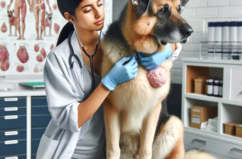
Chłoniak to jeden z najczęściej występujących nowotworów u psów, atakujący układ limfatyczny, w tym węzły chłonne, śledzionę i szpik kostny. Rak ten może pojawić się w różnych częściach ciała psa, często prowadząc do poważnych problemów zdrowotnych. Zrozumienie, jak wspierać i opiekować się psem chorym na chłoniaka, ma kluczowe znaczenie dla poprawy jakości jego życia. W tym artykule omówiono skuteczne strategie pomocy psom chorym na chłoniaka, koncentrując się zarówno na konwencjonalnych metodach leczenia, jak i możliwościach opieki wspomagającej.
Zrozumienie chłoniaka psów
Chłoniak u psów to rodzaj nowotworu wywodzącego się z limfocytów – komórek wchodzących w skład układu odpornościowego. Często wykrywa się ją na skutek powiększonych węzłów chłonnych, które można wyczuć pod skórą w takich miejscach jak szyja i za kolanami. Objawy mogą również obejmować letarg, utratę apetytu i niewyjaśnioną utratę wagi. Diagnozowanie chłoniaka zazwyczaj obejmuje biopsję węzłów chłonnych lub innych dotkniętych obszarów.
Konwencjonalne metody leczenia chłoniaka u psów
Podstawową metodą leczenia chłoniaka u psów jest chemioterapia, która w wielu przypadkach okazała się skuteczna. Konkretny protokół i czas trwania leczenia mogą się różnić w zależności od stadium i agresywności nowotworu. W niektórych przypadkach można również rozważyć radioterapię i operację, zwłaszcza jeśli guz jest zlokalizowany.
Opieka wspomagająca dla psów chorych na chłoniaka
Oprócz leczenia medycznego w leczeniu chłoniaka u psów niezbędne jest zapewnienie opieki wspomagającej. Oto kilka kluczowych strategii:
- Wsparcie żywieniowe: Kluczowa jest dobrze zbilansowana dieta dostosowana do potrzeb pacjenta chorego na nowotwór. Generalnie zaleca się spożywanie wysokiej jakości białka, zdrowych tłuszczów i ograniczonej ilości węglowodanów prostych w celu wsparcia układu odpornościowego i ogólnego stanu zdrowia.
- Zarządzanie bólem: Psy chore na chłoniaka mogą odczuwać ból, szczególnie w zaawansowanych stadiach. Leczenie bólu, które może obejmować przepisane leki przeciwbólowe i przeciwzapalne, ma kluczowe znaczenie dla utrzymania komfortu.
- Regularne monitorowanie: Częste kontrole u lekarza weterynarii są ważne w celu oceny skuteczności leczenia i wprowadzenia niezbędnych zmian. Monitorowanie pomaga również wcześnie wykryć wszelkie komplikacje.
- Wsparcie emocjonalne: Psy są bardzo wrażliwe na emocje swoich opiekunów. Zapewnienie spokojnego i pełnego miłości środowiska może pomóc zmniejszyć stres i poprawić ogólne samopoczucie.
- Terapie alternatywne: Niektórzy właściciele zwierząt domowych sięgają po terapie uzupełniające, takie jak akupunktura, masaż lub suplementy ziołowe, aby poprawić komfort i dobre samopoczucie. Ważne jest, aby omówić te opcje z lekarzem weterynarii, aby upewnić się, że są bezpieczne i potencjalnie korzystne.
Znaczenie wczesnego wykrywania
Wczesne wykrycie chłoniaka może znacząco wpłynąć na skuteczność leczenia. Właściciele zwierząt powinni regularnie sprawdzać swoje psy pod kątem objawów obrzęku lub guzków i zasięgnąć porady weterynaryjnej, jeśli zauważą jakiekolwiek nietypowe objawy.
Opieka nad psem chorym na chłoniaka obejmuje wieloaspektowe podejście, które obejmuje konwencjonalne leczenie raka i kompleksową opiekę wspomagającą. Rozumiejąc potrzeby swoich psich towarzyszy i ścisłą współpracę z lekarzami weterynarii, właściciele zwierząt mogą znacznie poprawić jakość życia swoich psów chorych na chłoniaka.

utworzone przez TCMVET | 18 maja 2024 r | Rak i guzy u psów
Chłoniak jest jednym z najpowszechniejszych typów nowotworów u psów, atakującym przede wszystkim układ limfatyczny, w tym węzły chłonne, śledzionę i szpik kostny. Nowotwór ten może objawiać się w różnych częściach ciała psa, stanowiąc poważne wyzwanie zdrowotne. Ponieważ właściciele zwierząt domowych coraz częściej poszukują łagodniejszych opcji leczenia, homeopatia stała się podejściem uzupełniającym. W tym miejscu zagłębiamy się w sposób, w jaki można zintegrować leczenie homeopatyczne z leczeniem chłoniaka u psów, podkreślając jego potencjalne korzyści i rozważania.
Zrozumienie chłoniaka u psów
Zanim zagłębisz się w leki homeopatyczne, ważne jest, aby zrozumieć, z czym wiąże się chłoniak. Ten typ nowotworu charakteryzuje się szybką proliferacją złośliwych limfocytów, które są rodzajem białych krwinek. Objawy mogą być bardzo zróżnicowane, ale często obejmują obrzęk węzłów chłonnych, letarg, utratę apetytu i utratę wagi. Tradycyjne metody leczenia zazwyczaj obejmują chemioterapię, która może być skuteczna, ale także dotkliwa, co prowadzi wiele osób do poszukiwania łagodniejszych alternatyw, takich jak homeopatia.
Homeopatyczne podejście do chłoniaka u psów
Homeopatia działa na zasadzie „podobne leczy podobne”, używając bardzo rozcieńczonych substancji w celu uruchomienia naturalnych procesów gojenia organizmu. Jest on dostosowywany indywidualnie do każdej osoby na podstawie jej specyficznych objawów i ogólnego temperamentu. W przypadku chłoniaka homeopata może wybrać środki mające na celu wzmocnienie układu odpornościowego, opanowanie objawów i poprawę jakości życia.
Typowe leki homeopatyczne na raka u psów
- Album Arsen: Często stosowany u psów wykazujących oznaki osłabienia, niepokoju i nadmiernego pragnienia.
- Calcarea Carbonica: Odpowiedni dla psów ospałych i mających tendencję do odczuwania zimna.
- Konium Maculatum: Stosowany w przypadkach, gdy występuje zauważalne stwardnienie i obrzęk gruczołów.
- Fosfor: Zalecany dla psów ze skłonnością do krwawień i tych, które potrzebują wzmocnienia odporności.
- Siarka: Dobry do poprawy ogólnej witalności, szczególnie jeśli pies ma problemy z obwisłym ciałem i skórą.
Integracja homeopatii z konwencjonalnymi metodami leczenia
Chociaż homeopatię można stosować samodzielnie, często służy ona jako uzupełnienie konwencjonalnych metod leczenia raka. Na przykład zintegrowanie homeopatii z chemioterapią może pomóc złagodzić skutki uboczne tradycyjnych leków i poprawić ogólne samopoczucie psa. Jednakże niezbędna jest konsultacja z lekarzem weterynarii mającym doświadczenie zarówno w medycynie konwencjonalnej, jak i holistycznej, aby opracować kompleksowy plan leczenia.
Uwagi i przestrogi
Ważne jest, aby podchodzić do leków homeopatycznych ze zrównoważonej perspektywy. Chociaż wielu właścicieli zwierząt domowych zgłasza poprawę zdrowia i dobrego samopoczucia swoich zwierząt dzięki homeopatii, dowody naukowe potwierdzające jej skuteczność, szczególnie w leczeniu raka, są nadal nieliczne. Zawsze omawiaj nowe leczenie z wykwalifikowanym lekarzem weterynarii, aby upewnić się, że pasuje ono do ogólnej strategii zdrowotnej Twojego psa.
Homeopatia oferuje obiecujące, uzupełniające podejście do leczenia chłoniaka u psów, koncentrując się na zindywidualizowanym leczeniu i substancjach naturalnych. Jak w przypadku każdej choroby, zwłaszcza nowotworu, kluczowa jest współpraca z lekarzem weterynarii. Niezależnie od tego, czy jest to samodzielna terapia, czy dodatek do metod konwencjonalnych, homeopatia może potencjalnie poprawić jakość życia i zdrowie psów chorych na chłoniaka, torując drogę do holistycznego powrotu do zdrowia.

utworzone przez TCMVET | 17 maja 2024 r | Rak i guzy u psów
Rozwój nowotworu u psów może być niepokojącym problemem dla każdego właściciela zwierzęcia. Zrozumienie, jak zapobiegać lub spowalniać rozwój guza, może znacząco poprawić jakość życia psa i wydłużyć jego długość życia. W artykule omówiono kompleksowe strategie łączące środki zapobiegawcze i skuteczne techniki leczenia w celu zwalczania wzrostu guza u psów.
1. Regularne kontrole weterynaryjne
Wczesne wykrycie jest kluczem do skutecznego kontrolowania wzrostu nowotworu u psów. Regularne kontrole weterynaryjne, najlepiej dwa razy w roku w przypadku dorosłych psów i częściej w przypadku seniorów, pozwalają na wczesną identyfikację i leczenie wszelkich podejrzanych narośli, zanim się rozwiną. Kontrole te powinny obejmować dokładne badania fizykalne i, jeśli to konieczne, diagnostykę obrazową, taką jak prześwietlenia rentgenowskie lub ultradźwięki.
2. Prawidłowe odżywianie
Karmienie psa zbilansowaną dietą wysokiej jakości ma kluczowe znaczenie w zapobieganiu nowotworom. Diety bogate w przeciwutleniacze, takie jak witaminy A, C i E, mogą pomóc chronić komórki przed uszkodzeniami i zmniejszyć ryzyko raka. Włączaj świeże, pełnowartościowe pokarmy, takie jak chude mięso, zdrowe tłuszcze, takie jak olej rybny, i warzywa, aby wspierać ogólny stan zdrowia i funkcje odpornościowe.
3. Utrzymuj zdrową wagę
Otyłość jest znanym czynnikiem ryzyka różnych typów nowotworów. Utrzymanie prawidłowej wagi psa nie tylko zmniejsza ryzyko rozwoju nowotworu, ale także pomaga w ogólnym zdrowiu i witalności. Regularne ćwiczenia i kontrola porcji to istotne elementy kontroli wagi.
4. Minimalizuj narażenie na czynniki rakotwórcze
Ograniczenie narażenia psa na czynniki rakotwórcze może pomóc w zapobieganiu wystąpieniu nowotworów. Unikaj biernego palenia, chemii do trawników i szkodliwych domowych środków czyszczących. Wybieraj produkty naturalne zarówno w domu, jak i na podwórku, aby chronić środowisko swojego zwierzaka tak bardzo, jak to możliwe.
5. Sterylizacja lub kastracja
Sterylizacja lub kastracja psa może znacznie zmniejszyć ryzyko wystąpienia niektórych rodzajów nowotworów, szczególnie tych związanych z układem rozrodczym, takich jak nowotwory sutka u kobiet i rak jąder u mężczyzn. Skonsultuj się ze swoim lekarzem weterynarii w sprawie najlepszego wieku do przeprowadzenia tych zabiegów, ponieważ czas może mieć wpływ na ich działanie ochronne przed rakiem.
6. Stosowanie immunoterapii i suplementów
Pojawiające się terapie, takie jak immunoterapia, okazują się obiecujące, pomagając układowi odpornościowemu rozpoznawać i zwalczać komórki nowotworowe u psów. Ponadto suplementy diety, takie jak kurkuma, która zawiera kurkuminę, mają właściwości przeciwzapalne i przeciwnowotworowe, które mogą pomóc w spowolnieniu wzrostu guza.
7. Regularna opieka stomatologiczna
Zły stan zębów może być ukrytym źródłem przewlekłego stanu zapalnego, który może przyczyniać się do rozwoju raka. Regularne kontrole i czyszczenie zębów, a także codzienne szczotkowanie zębów są niezbędne do utrzymania zdrowia jamy ustnej psa i potencjalnie zmniejszają ryzyko nowotworów jamy ustnej.
8. Redukcja stresu
Przewlekły stres może osłabić układ odpornościowy i potencjalnie zwiększyć ryzyko wzrostu nowotworu. Zapewnij stabilne i pełne miłości środowisko domowe, regularne ćwiczenia i stymulację umysłową, aby pomóc Twojemu psu skutecznie radzić sobie ze stresem.
Stosując te proaktywne strategie, możesz znacząco wpłynąć na ryzyko rozwoju nowotworu u psa i zarządzanie nim, co doprowadzi do zdrowszego i szczęśliwszego życia Twojego futrzanego przyjaciela.

utworzone przez TCMVET | 17 maja 2024 r | Rak i guzy u psów
Rak u psów, podobnie jak u ludzi, może prowadzić do poważnych problemów, w tym utraty wagi, co może mieć wpływ na ogólny stan zdrowia i samopoczucie Twojego zwierzaka. Skuteczne zarządzanie utratą wagi jest kluczowe, ponieważ może poprawić jakość życia psa, zwiększyć jego poziom energii i potencjalnie poprawić jego reakcję na leczenie raka. W tym artykule omawiamy praktyczne i zalecane przez weterynarza strategie, które pomogą Twojemu psiemu towarzyszowi przybrać na wadze podczas walki z rakiem.
1. Skonsultuj się ze swoim lekarzem weterynarii
Przed wprowadzeniem jakichkolwiek zmian w diecie lub sposobie pielęgnacji psa należy skonsultować się z lekarzem weterynarii. Mogą zapewnić dostosowany plan w oparciu o konkretny rodzaj raka Twojego psa, aktualny protokół leczenia i ogólny stan zdrowia. Ten krok jest kluczowy, aby upewnić się, że jakiekolwiek zmiany w diecie nie zakłócają leczenia.
2. Wysokokaloryczna i bogata w składniki odżywcze żywność
Psy chore na raka potrzebują wysokokalorycznej i bogatej w składniki odżywcze diety, aby pomóc utrzymać wagę. Weź pod uwagę produkty bogate w białko i tłuszcze, które są niezbędne do utrzymania energii i masy ciała. Twój weterynarz może zalecić dietę na receptę przygotowaną specjalnie dla psów chorych na raka. Diety te są opracowane tak, aby były wysoce strawne i atrakcyjne, aby zachęcać do jedzenia pomimo zmniejszonego apetytu.
3. Częste, małe posiłki
Zamiast dwóch dużych posiłków, oferuj mniejsze, ale częstsze posiłki w ciągu dnia. Mniejsze posiłki są łatwiejsze do strawienia i mogą zmniejszyć obciążenie układu trawiennego psa. Może to również pomóc w utrzymaniu stałego poziomu energii przez cały dzień.
4. Stymulanty apetytu
Jeśli Twój pies nie wykazuje zainteresowania jedzeniem, lekarz weterynarii może przepisać środki pobudzające apetyt. Leki te mogą pomóc zwiększyć chęć jedzenia u psa, co jest szczególnie pomocne, jeśli pies przechodzi chemioterapię lub inne leczenie, które może zmniejszyć jego apetyt.
5. Smaczna i miękka żywność
Czasami rak i jego leczenie mogą sprawić, że jedzenie będzie dla psów niewygodne. Oferowanie smacznej, miękkiej lub mokrej żywności może zachęcić je do jedzenia więcej. Możesz także podgrzać jedzenie, aby poprawić jego zapach i uczynić go bardziej atrakcyjnym.
6. Suplementy diety
Porozmawiaj ze swoim weterynarzem o możliwości włączenia suplementów diety do diety Twojego psa. Suplementy, takie jak olej rybny, bogaty w kwasy tłuszczowe omega-3, mogą pomóc w walce z utratą wagi i zapewnić niezbędne kalorie i składniki odżywcze, których potrzebuje Twój pies.
7. Zapewnij im wygodę i bezstresowość
Komfortowe otoczenie może pomóc Twojemu psu poczuć się bardziej zrelaksowanym i chętnym do jedzenia. Upewnij się, że miejsce do spożywania posiłków jest ciche i z dala od domowego hałasu i stresu. Komfort może znacząco wpływać na apetyt i zachowania żywieniowe.
8. Monitoruj postępy swojego psa
Regularnie monitoruj wagę psa i jego nawyki żywieniowe. Prowadź dziennik dziennego spożycia pokarmu i zmian masy ciała i udostępniaj te informacje swojemu lekarzowi weterynarii. Pomoże to w dostosowaniu ich planu żywieniowego w razie potrzeby, aby upewnić się, że podążają właściwą drogą.
Kontrola masy ciała u psów chorych na raka to delikatna kwestia, która wymaga dbałości o szczegóły i ścisłej współpracy z lekarzem weterynarii. Stosując te strategie, możesz pomóc swojemu psu nie tylko utrzymać, ale potencjalnie przybrać na wadze, przyczyniając się do jego siły i witalności podczas walki z rakiem.

utworzone przez TCMVET | 15 maja 2024 r | Rak i guzy u psów
Jeśli chodzi o zarządzanie zdrowiem psa, szczególnie w przypadku raka lub guzów tłuszczowych, proaktywne podejście może mieć znaczące znaczenie. W tym artykule omówiono skuteczne suplementy zwalczające raka i praktyczne metody zmniejszania guzów tłuszczowych u psów, oferując właścicielom zwierząt cenne informacje, które mogą pomóc poprawić samopoczucie ich futrzanych przyjaciół.
Suplementy zwalczające raka dla psów
Włączenie określonych suplementów do diety psa może wspierać jego układ odpornościowy i potencjalnie hamować rozwój raka. Oto kilka powszechnie zalecanych suplementów:
- Kwasy tłuszczowe omega-3: Kwasy tłuszczowe omega-3, występujące obficie w oleju rybnym, mogą pomóc w zmniejszeniu stanu zapalnego, co jest kluczowe, ponieważ przewlekłe zapalenie może przyczyniać się do progresji raka.
- Kurkuma (Curcumin): Znana ze swoich właściwości przeciwzapalnych, kurkumina ma również działanie przeciwnowotworowe. Pomaga w spowolnieniu rozprzestrzeniania się komórek nowotworowych i zmniejszeniu stanu zapalnego.
- Ostropest plamisty: Zioło to wspomaga zdrowie wątroby i jest szczególnie korzystne dla psów poddawanych chemioterapii, ponieważ pomaga chronić przed toksycznością wątroby.
- Ekstrakty z grzybów: Niektóre grzyby, takie jak ogon indyczy, zawierają polisacharydy, które wzmacniają układ odpornościowy i są powiązane z leczeniem raka u ludzi i zwierząt.
Przed rozpoczęciem stosowania nowego suplementu skonsultuj się z weterynarzem, który może udzielić wskazówek w oparciu o specyficzne potrzeby zdrowotne Twojego psa i istniejące metody leczenia.
Strategie zmniejszania guzów tłuszczowych u psów
Guzy tłuszczowe, czyli tłuszczaki, są częste u psów, szczególnie w starszym wieku. Chociaż są one zazwyczaj łagodne, zmniejszenie ich rozmiaru może pomóc w utrzymaniu komfortu i mobilności psa. Oto kilka strategii do rozważenia:
- Ulepszona dieta: Zmniejszenie spożycia kalorii i skupienie się na diecie bogatej w błonnik i niskotłuszczowej może pomóc w kontrolowaniu wielkości guzów tłuszczowych.
- Regularne ćwiczenia: Aktywność psa pomaga spalać tłuszcz, co może bezpośrednio wpływać na wielkość i rozwój guzów tłuszczowych.
- Naturalne suplementy: Oprócz suplementów zwalczających raka, niektóre naturalne substancje, takie jak olej lniany, który jest bogaty w kwasy omega-3, mogą pomóc w zmniejszeniu rozmiaru tłuszczaka poprzez promowanie metabolizmu tłuszczów.
Wdrażanie Planu
Włączenie tych suplementów i strategii do rutyny Twojego psa wymaga zrównoważonego podejścia. Regularne wizyty kontrolne u lekarza weterynarii pozwolą monitorować skuteczność wybranych metod i w razie potrzeby korygować plan. Celem jest opanowanie objawów i poprawa jakości życia, a nie tylko skupianie się na chorobie lub nowotworach.
Leczenie raka i guzów tłuszczowych u psów obejmuje kompleksową strategię obejmującą odpowiednie suplementy i zmiany stylu życia. Stosując te praktyki, właściciele zwierząt mogą odegrać aktywną rolę we wspieraniu zdrowia swoich psów, potencjalnie przedłużając ich życie i poprawiając ich codzienny komfort.

utworzone przez TCMVET | 15 maja 2024 r | Rak i guzy u psów
Rak u psów to trudna diagnoza dla każdego właściciela zwierzęcia. Podczas gdy tradycyjna medycyna weterynaryjna oferuje chirurgię, chemioterapię i radioterapię, wielu właścicieli zwierząt domowych zwraca się również w stronę podejścia holistycznego, takiego jak leki homeopatyczne, aby zapewnić komfort i prawdopodobnie wydłużyć jakość życia swoich psich towarzyszy. W tym artykule zbadano integrację terapii homeopatycznych ze strategiami opieki wspomagającej, aby pomóc psom, u których zdiagnozowano raka.
Zrozumienie homeopatycznego leczenia raka psów
Homeopatia jest formą medycyny alternatywnej opartą na zasadzie „podobne leczy podobne”, która sugeruje, że substancje wywołujące objawy u zdrowego człowieka mogą leczyć podobne objawy u chorego, jeśli są podawane w silnie rozcieńczonych dawkach. W przypadku psów chorych na raka leki homeopatyczne są dostosowywane do indywidualnych objawów, a nie do samej choroby.
Typowe leki homeopatyczne stosowane w leczeniu raka u psów obejmują:
- Album Arsen: Często stosowany u zwierząt doświadczających poważnego osłabienia, niepokoju i niepokoju.
- Thuja zachodnia: Generalnie zalecany w przypadku odrostów wynikających ze szczepień lub innych podstawowych problemów.
- Belladona: Odpowiedni w przypadkach, w których objawy pojawiają się nagle, są szczególnie intensywne i bolesne.
Bardzo ważne jest, aby skonsultować się z weterynarzem specjalizującym się w homeopatii, aby wybrać odpowiedni lek i dawkowanie dla konkretnego stanu Twojego psa.
Strategie opieki wspomagającej dla psów chorych na raka
Oprócz leczenia homeopatycznego, opieka wspomagająca jest niezbędna w kontrolowaniu jakości życia psa chorego na raka. Oto kilka skutecznych strategii opieki wspomagającej:
- Odżywianie: Karmienie psa zbilansowaną dietą dostosowaną do jego specyficznych potrzeb może pomóc w utrzymaniu siły i poprawie jakości życia. Niektórzy właściciele zwierząt decydują się na dietę bogatą w przeciwutleniacze, kwasy tłuszczowe omega-3 i łatwo przyswajalne białka.
- Zarządzanie bólem: Tradycyjne leki przeciwbólowe, akupunktura i laseroterapia to opcje pomagające skutecznie radzić sobie z bólem.
- Regularne ćwiczenia: W zależności od zdrowia i wytrzymałości psa, lekkie lub umiarkowane ćwiczenia mogą pomóc w utrzymaniu napięcia mięśniowego i pobudzić jego ducha.
- Wsparcie emocjonalne: Podobnie jak ludzie, psy czerpią korzyści ze wsparcia i miłości w środowisku. Utrzymanie rutyny i wspólne spędzanie czasu może pomóc podnieść psa na duchu.
Uwagi i środki ostrożności
Chociaż leki homeopatyczne oferują alternatywną ścieżkę, konieczne jest kompleksowe podejście do leczenia raka. Zawsze omawiaj wszelkie nowe plany leczenia z wykwalifikowanym lekarzem weterynarii, który rozumie zarówno podejście tradycyjne, jak i holistyczne. Monitorowanie reakcji psa na leczenie i dostosowywanie go w razie potrzeby ma kluczowe znaczenie.
Podsumowując, leczenie raka u psów wymaga połączenia interwencji medycznej i troskliwej opieki. Niezależnie od tego, czy integrujesz homeopatię, dostosowujesz potrzeby dietetyczne, czy zapewniasz wsparcie emocjonalne, celem pozostaje poprawa jakości życia Twojego psa i zapewnienie najlepszej możliwej opieki podczas jego leczenia.


