
oleh TCMVET | 23 Sep 2024 | Kanker & Tumor Anjing
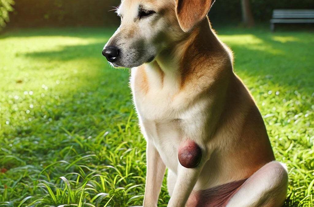
Apocrine gland cysts and adenomas are common benign conditions seen in middle-aged to older dogs, particularly affecting breeds like the Great Pyrenees, Chow Chows, and Alaskan Malamutes. These cysts and adenomas develop from apocrine glands, which are associated with hair follicles and are found in the skin. This article provides an in-depth look at these conditions, including causes, symptoms, and treatment options.
What are Apocrine Gland Cysts?
Apocrine gland cysts are non-cancerous cysts that occur in the skin of dogs. They are typically seen in the middle to upper skin layers, loosely associated with hair follicles. Apocrine cysts can form on the head, neck, and other areas, often presenting as one or more fluid-filled sacs under the skin. These cysts are usually benign, meaning they do not spread or become cancerous.
There are two forms of apocrine gland cysts:
- Localized Apocrine Cysts: These develop in or near individual hair follicles and are more commonly found on the head and neck of dogs.
- Diffuse Apocrine Cysts: This form involves multiple cysts forming in the apocrine glands associated with multiple hair follicles. They can appear in areas of uninjured skin, making them harder to treat if they become widespread.
While benign, these cysts may sometimes cause discomfort, especially if they grow large or rupture.
What are Apocrine Gland Adenomas?
Apocrine gland adenomas are benign tumors that can arise in the apocrine glands. They appear as firm to soft masses and may contain fluid that ranges in color from clear to brownish. Typically, these cysts are not larger than 1.6 inches (4 centimeters) in diameter. Like apocrine cysts, adenomas are also found on the head, neck, and legs of older dogs, and, in rare cases, cats and horses.
Apocrine adenomas come in two types:
- Apocrine Adenomas: These are solid, benign growths formed by the apocrine glands.
- Apocrine Ductular Adenomas: These involve the ducts of the apocrine glands, leading to cystic formations.
Breeds at Higher Risk
Certain dog breeds are more prone to developing apocrine gland cysts and adenomas. The most commonly affected breeds include:
- Pyrenees Besar
- Chow Chows
- Malamute Alaska
Older dogs in these breeds are more likely to develop these conditions, though the exact cause remains unknown. Genetics, environmental factors, and age may play a role in their development.
Gejala yang Harus Diperhatikan
- Visible cysts or lumps on the head, neck, or legs
- Fluid-filled sacs under the skin
- Soft to firm masses that may vary in size
- Discoloration of the skin or cyst fluid (clear to brown)
- Possible rupture of cysts causing localized inflammation or infection
While these cysts and adenomas are generally painless, they can cause discomfort if they rupture or grow in sensitive areas.
Pilihan pengobatan
The primary treatment for apocrine gland cysts and adenomas is surgical removal. However, this can be challenging, particularly in cases where cysts are diffuse and spread across a larger area of the skin. In localized cases, the surgery is typically straightforward, and the prognosis is excellent after removal.
In situations where surgery is not an option, veterinarians may recommend monitoring the cysts for changes in size or discomfort. If cysts rupture, antibiotics may be required to prevent infection.
Apocrine gland cysts and adenomas in dogs are generally benign and treatable conditions. While they can cause cosmetic issues and occasional discomfort, early detection and treatment can help prevent complications. Owners of at-risk breeds such as Great Pyrenees, Chow Chows, and Alaskan Malamutes should monitor their dogs for the development of these cysts as they age, and consult a veterinarian for proper diagnosis and treatment.

oleh TCMVET | 22 Sep 2024 | Kanker & Tumor Anjing
Cornifying epitheliomas, also known as keratoacanthomas or infundibular keratinizing acanthomas, are benign skin tumors that commonly affect middle-aged dogs. These tumors are characterized by tough, layered lumps that protrude from the skin and can resemble small horns. While generally harmless, they may cause discomfort or lead to complications if left untreated. This article explores the causes, symptoms, and treatment options for cornifying epitheliomas in dogs.
What Are Cornifying Epitheliomas?
Cornifying epitheliomas are benign tumors that typically arise from hair follicles. These growths form nests of tough, layered lumps on the skin, which may look like small horns or cornified cysts. They can develop anywhere on the dog’s body but are most frequently found on the back, tail, and legs. In some cases, these tumors may cause irritation, leading to scratching or biting, which can result in secondary infections or skin trauma.
Breeds at Risk for Cornifying Epitheliomas
Certain dog breeds are more prone to developing cornifying epitheliomas. Middle-aged dogs are particularly susceptible, and specific breeds at higher risk include:
- Norwegian Elkhounds
- Anjing Gembala Belgia
- Lhasa Apsos
- Bearded Collies
Norwegian Elkhounds and Lhasa Apsos are particularly at risk for developing multiple tumors or a more widespread form of the disease.
Symptoms of Cornifying Epitheliomas
The primary symptom of cornifying epitheliomas is the presence of tough, horn-like tumors on the skin. These tumors can vary in size and may cause discomfort depending on their location. Some key symptoms include:
- Raised, horn-like lumps: The tumors appear as tough, layered growths that stick up from the skin surface.
- Cornified cysts: In some cases, the tumors may appear as cornified cysts rather than horns.
- Self-trauma: Dogs may scratch, rub, or bite at the tumors, leading to trauma or ulceration.
- Secondary infections: If the tumors are irritated or broken open, they can become infected.
Causes of Cornifying Epitheliomas
While the exact cause of cornifying epitheliomas is not fully understood, they most likely originate from hair follicles. Genetic factors may play a role in certain breeds, especially those predisposed to the condition. Environmental factors such as skin irritation or trauma may also contribute to the development of these tumors.
Treatment Options for Cornifying Epitheliomas
Treatment for cornifying epitheliomas depends on the severity of the condition and whether the tumors are causing discomfort or complications. In some cases, the tumors may be left untreated if they are not causing any issues. However, treatment is recommended in cases of self-trauma, ulceration, or secondary infection.
- Operasi pengangkatan: Surgical removal is the preferred treatment option, especially if the dog is experiencing discomfort or if the tumors are prone to infection. However, it’s important to note that dogs are likely to develop additional tumors over time.
- Oral Retinoid Medications: For dogs with a generalized form of the disease, oral retinoid medications may be prescribed to help manage the condition and reduce the occurrence of new tumors.
- Pemantauan: In cases where the tumors are not causing any issues, regular monitoring is advised to ensure they do not worsen or lead to complications.
Cornifying epitheliomas are benign tumors that can cause discomfort and complications in dogs, particularly in certain breeds like Norwegian Elkhounds and Lhasa Apsos. While treatment is not always necessary, surgical removal is recommended in cases where the tumors lead to self-trauma or infection. With proper management, most dogs can live comfortably with this condition, although additional tumors may develop over time.

oleh TCMVET | 22 Sep 2024 | Kanker & Tumor Anjing
Cutaneous angiosarcomas, also known as angioendotheliomas, are malignant tumors that arise from blood vessels in the skin. These tumors can initially resemble benign hemangiomas but later progress into aggressive malignancies. While they can affect many breeds, dogs with short, white coats and high exposure to sunlight are particularly susceptible. In this article, we will explore the causes, symptoms, and treatment options for cutaneous angiosarcomas in dogs.
What Are Cutaneous Angiosarcomas?
Cutaneous angiosarcomas are rare but aggressive tumors that develop from the blood vessels in the skin. These tumors can appear on various parts of the body, most often affecting the underside of the trunk, hips, thighs, and lower legs in dogs. While they may initially appear harmless, resembling benign hemangiomas, they can become malignant and spread to surrounding tissues.
Breeds Prone to Angiosarcomas
Certain dog breeds are more susceptible to cutaneous angiosarcomas, either due to their coat type or genetic predisposition.
- Sun-Related Risk: Dogs with short, white coats, such as Whippets, Italian Greyhounds, white Boxers, Dan Pit Bull Terriers, are more likely to develop sun-caused angiosarcomas due to their increased exposure to ultraviolet (UV) radiation.
- Genetic Risk: Breeds like Wolfhound Irlandia, Vizslas, Anjing Golden Retriever, Dan Gembala Jerman are also prone to developing these tumors, although not as a result of sun exposure.
Causes of Cutaneous Angiosarcomas
Sun exposure is a significant risk factor for developing cutaneous angiosarcomas in breeds with short, light-colored coats. Prolonged UV radiation can damage the skin’s cells, leading to mutations that eventually result in tumor formation. In non-sun-related cases, genetic factors likely contribute to the development of angiosarcomas in predisposed breeds.
Symptoms of Cutaneous Angiosarcomas
Cutaneous angiosarcomas may initially present as small, benign-looking lumps on the skin. Over time, these tumors may grow and become more aggressive. Common symptoms include:
- Red or purple bumps: These bumps often resemble bruises or benign hemangiomas.
- Koreng: As the tumor progresses, it may break open and ulcerate, causing discomfort and infection.
- Pembengkakan: Localized swelling in the affected area may occur as the tumor grows.
- Pertumbuhan cepat: These tumors can grow quickly, spreading to nearby tissues.
Treatment Options for Cutaneous Angiosarcomas
The treatment for cutaneous angiosarcomas largely depends on the size and location of the tumor. Early detection and intervention are crucial for successful management. Treatment options include:
- Bedah Krios: Small surface tumors can often be treated with freezing, a procedure known as cryosurgery. This method effectively destroys the tumor cells with minimal invasiveness.
- Bedah untuk FOSCC terutama berfokus pada reseksi tumor—baik pengangkatan parsial atau lengkap dari tumor.: Laser surgery can also be used to remove small tumors, offering a precise and effective treatment option with a lower risk of scarring.
- Sun Exposure Avoidance: For dogs prone to sun-related angiosarcomas, reducing UV exposure is critical. Limiting time in the sun, applying dog-safe sunscreen, and using protective clothing can help prevent new tumors from forming.
- Pemantauan Jangka Panjang: Even after successful treatment, new tumors may develop over time, so long-term monitoring is essential to catch any future growths early.
Cutaneous angiosarcomas are aggressive blood vessel tumors that can significantly impact a dog’s health, particularly for breeds with high sun exposure or genetic predispositions. Early detection and prompt treatment are vital for managing the condition. Pet owners can help reduce their dog’s risk by minimizing sun exposure and seeking veterinary advice at the first sign of any suspicious skin growths.

oleh TCMVET | 22 Sep 2024 | Kanker & Tumor Anjing
Hamartoma epidermal, juga dikenal sebagai nevi, adalah kondisi kulit langka yang muncul sebagai benjolan gelap dan runcing di kulit anjing. Meskipun mereka jinak, pertumbuhan ini dapat menyebabkan masalah kosmetik dan rentan terhadap infeksi sekunder, terutama pada anak anjing. Dalam artikel ini, kita akan menjelajahi gejala, penyebab, dan opsi pengobatan untuk hamartoma epidermal pada anjing.
Apa Itu Hamartoma Epidermal?
Hamartoma epidermal adalah lesi kulit jinak yang membentuk benjolan gelap dan terangkat, terkadang tersusun dalam garis di kulit. Benjolan ini biasanya lebih umum pada anjing muda dan anak anjing, dan meskipun mereka mungkin terlihat tidak menyenangkan, mereka tidak bersifat kanker. Namun, hamartoma dapat membentuk jerawat atau lipatan kulit yang menebal, yang menyebabkan ketidaknyamanan dan potensi infeksi.
Gejala Hamartoma Epidermal
Tanda utama hamartoma epidermal adalah munculnya benjolan gelap dan runcing di kulit anjing. Pertumbuhan ini dapat bervariasi dalam ukuran dan bentuk serta dapat muncul dalam berbagai bentuk:
- Benjolan runcing dan gelap: Nevi mungkin kecil dan gelap, muncul sebagai pertumbuhan runcing di permukaan kulit.
- Jerawat atau lipatan kulit yang tebal: Dalam beberapa kasus, kulit di sekitar hamartoma menebal atau membentuk struktur mirip jerawat.
- Susunan linier: Kadang-kadang, benjolan ini muncul dalam garis, yang bisa menjadi ciri khas dari kondisi ini.
- Rentan terhadap infeksi: Karena sifat pertumbuhan ini, mereka dapat menjebak bakteri, yang menyebabkan infeksi sekunder, terutama jika anjing menggaruk atau mengiritasi area tersebut.
Penyebab dan Faktor Risiko
Meskipun penyebab pasti hamartoma epidermal tidak selalu diketahui, beberapa faktor berkontribusi pada perkembangannya:
- Genetics: Pada ras tertentu, seperti Cocker Spaniel, hamartoma epidermal dapat diwariskan. Predisposisi genetik ini membuat mereka lebih mungkin mengembangkan kondisi ini pada usia muda.
- Anak Anjing: Pertumbuhan kulit ini lebih sering diamati pada anak anjing, meskipun dapat muncul pada anjing dari segala usia.
Opsi Pengobatan untuk Hamartoma Epidermal
Pengobatan hamartoma epidermal sebagian besar tergantung pada ukuran dan jumlah pertumbuhan. Meskipun mereka jinak, risiko infeksi dan penampilan mereka yang tidak sedap dipandang sering mendorong pengobatan. Opsi termasuk:
- Operasi pengangkatan: Hamartoma kecil biasanya dapat diangkat secara bedah dengan komplikasi minimal. Ini sering menjadi opsi yang diutamakan jika nevi sedikit dan terlokalisasi.
- Pengobatan obat: Untuk anjing dengan hamartoma besar atau banyak, pengangkatan bedah mungkin tidak memungkinkan. Dalam kasus ini, pengobatan dapat membantu mengelola kondisi tersebut. Obat anti-inflamasi atau antibiotik mungkin diresepkan jika ada infeksi sekunder.
- Pemantauan rutin: Untuk lesi jinak yang tidak menyebabkan ketidaknyamanan atau infeksi, pemantauan rutin oleh dokter hewan mungkin cukup untuk memastikan pertumbuhan tidak memburuk.
Hamartoma epidermal jarang terjadi, kondisi kulit jinak yang paling umum terlihat pada anak anjing dan ras tertentu, seperti Cocker Spaniel. Meskipun tidak berbahaya, penampilannya dan risiko infeksi sering kali membuat pengangkatan atau pengobatan diperlukan. Jika Anda melihat benjolan atau perubahan kulit yang tidak biasa pada anjing Anda, konsultasikan dengan dokter hewan untuk menentukan langkah terbaik.
oleh TCMVET | 21 Sep 2024 | Kanker & Tumor Anjing
Basal cell tumors are one of the most common types of benign skin tumors found in dogs. These growths typically affect middle-aged to older dogs and can appear as firm, dome-shaped masses on the skin. While generally non-cancerous, basal cell tumors can still cause discomfort due to their size and the potential for ulceration. In this article, we will explore the symptoms, causes, and treatment options for basal cell tumors in dogs.
What Are Basal Cell Tumors?
Basal cell tumors are skin growths that originate from the basal cells found in a dog’s skin. These tumors are typically benign, meaning they are not cancerous, but they can grow large and cause discomfort. They are most commonly found on the dog’s head (especially the ears), neck, and forelimbs. Although they are benign, they may cause problems such as ulceration, inflammation, and discomfort, particularly if they grow in size.
Symptoms of Basal Cell Tumors in Dogs
Basal cell tumors generally present as firm, elevated masses on the skin. These growths may vary in size, from small bumps less than 0.4 inches (1 centimeter) to large masses over 4 inches (10 centimeters) in diameter. Some additional characteristics include:
- Solitary lumps: The tumors often appear as single masses rather than multiple growths.
- Hairless or ulcerated surface: These lumps are frequently hairless, and in some cases, they may break open and ulcerate.
- Dome-shaped growths: The masses tend to have a dome-like shape, often sticking out from the skin on stalk-like projections.
- Dark coloration: In some instances, the tumors can be dark in color.
- Secondary cysts: Cysts may form within or around the tumor.
Despite their benign nature, basal cell tumors can become problematic when they ulcerate and cause secondary inflammation. Dogs with these tumors may experience discomfort, especially if the mass becomes infected or leads to the death of surrounding skin tissue.
Causes of Basal Cell Tumors in Dogs
Several factors may contribute to the development of basal cell tumors in dogs:
- Usia: These tumors are more common in middle-aged to older dogs.
- Breed predisposition: Certain breeds, including Wirehaired Pointing Griffons, Kerry Blue Terriers, and Wheaten Terriers, are more likely to develop basal cell tumors.
- Paparan sinar matahari: Prolonged exposure to UV radiation may increase the risk of skin tumors in dogs.
Treatment Options for Basal Cell Tumors in Dogs
The most effective treatment for basal cell tumors is surgical removal. Since these tumors can grow large and cause discomfort due to ulceration and inflammation, removing the tumor eliminates the source of irritation. Surgery is typically straightforward, especially when the tumor is caught early before it grows too large.
In some cases, additional treatments such as antibiotics or anti-inflammatory medications may be prescribed to manage secondary infections or inflammation caused by the tumor. Once the tumor is removed, the chances of recurrence are generally low, and the dog’s quality of life improves significantly.
Basal cell tumors in dogs, while benign, can cause significant discomfort due to their size and potential for ulceration. Regular monitoring of your dog’s skin, especially if they belong to a breed predisposed to these tumors, can help catch any growths early. Surgical removal is an effective treatment and can prevent further complications such as infections or inflammation. If you notice any unusual lumps or masses on your dog’s skin, consult a veterinarian to determine the best course of action.

oleh TCMVET | 21 Sep 2024 | Kanker & Tumor Anjing
Skin tags, also known as acrochordons, are common benign skin lumps that frequently appear on older dogs. While harmless, these growths can sometimes cause concern for pet owners due to their appearance and the possibility of multiple occurrences. In this article, we will explore what skin tags are, why they develop, how they are diagnosed, and the available treatment options.
What Are Skin Tags in Dogs?
Skin tags are small, benign growths that can appear anywhere on a dog’s body. They often resemble stalk-like extensions and may have a wart-like surface. Although these growths can affect dogs of any breed, larger breeds tend to be at higher risk. Skin tags typically do not cause discomfort or pain unless they become irritated or injured.
Causes of Skin Tags in Dogs
While the exact cause of skin tags is not fully understood, several factors contribute to their development, particularly in older dogs. These factors include:
- Aging: As dogs age, their skin undergoes changes, making older dogs more susceptible to developing skin tags.
- Genetics: Certain breeds, especially large ones, are more prone to developing skin tags.
- Friction: Areas of the skin that experience frequent rubbing or friction, such as under the collar or around the legs, may be more likely to develop skin tags.
Diagnosing Skin Tags in Dogs
Skin tags are generally harmless, but it is essential to have them properly diagnosed by a veterinarian. This is particularly important as some skin conditions, such as warts or tumors, can appear similar to skin tags. A veterinarian may recommend a biopsi to confirm that the growth is benign. If your dog develops one skin tag, it is common for others to appear over time.
Treatment Options for Skin Tags in Dogs
While most skin tags do not require removal, surgical intervention may be considered if the growth becomes irritated, infected, or unsightly. Removal is typically done using methods such as excision, laser removal, or cryotherapy (freezing). However, unless the skin tag is causing discomfort or affecting the dog’s quality of life, surgery is often not necessary.
Skin tags in dogs are generally harmless but may cause worry for pet owners due to their appearance. Proper diagnosis and understanding of their benign nature can alleviate concerns. If a skin tag becomes problematic, consulting a veterinarian about removal options can help keep your dog comfortable and healthy.
