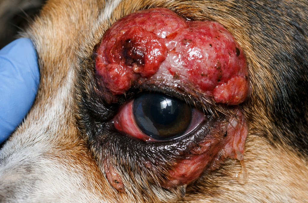
“Spindle cell” usually describes how tumor cells look under the microscope—long, tapered, and fibrous—rather than naming a single cancer type. In dogs, this description often overlaps with soft tissue sarcomas (STS) and related sarcoma-type tumors, which are known for local invasiveness: they can extend microscopically beyond what you can see or feel. That growth pattern is one big reason local recurrence becomes a key concern.
How rare is a spindle cell tumor near the eye?
It depends on whether the mass is a superficial eyelid growth or a deeper periocular/orbital tumor.
Many canine eyelid tumors are common and often benign, which is reassuring in everyday eye practice. However, a malignant spindle-cell/sarcoma-type tumor that is fast-growing or deep around the eye is a different scenario. Orbital and retrobulbar disease (disease in the eye socket or behind the globe) is less common than simple eyelid bumps and typically demands more complex planning.
Symptoms owners commonly notice
A tumor “over the eye” can look like a firm lump or swelling near the brow or eyelids, but deeper disease may show up through function changes. Dogs may squint, tear excessively, develop persistent discharge, rub at the eye or face, or seem head-shy. If the orbit is involved, classic signs can include exophthalmos (a bulging eye), third eyelid protrusion, and pain around the eye.
Pain and suffering: how much does it hurt?
Pain varies widely and is driven by what the tumor is doing locally.
A mass can become very uncomfortable if it distorts eyelid position or causes chronic corneal irritation with blinking. Veterinary ophthalmology guidance notes that eyelid masses often enlarge over time and can irritate the ocular surface, affecting comfort and quality of life. Orbital disease can also be painful—especially when swelling, pressure, or inflammation is present—and pain is frequently mentioned among the clinical signs in orbital conditions.
Why treatment near the eye is so challenging
For sarcoma-type tumors, the goal is usually strong local control, because microscopic tumor “fingers” can extend beyond what looks abnormal. On the trunk or limbs, surgeons often aim for wider margins to remove those invisible extensions. Around the eye, wide margins can be hard to achieve without damaging critical structures, which is why doctors may sound cautious about guarantees.
Treatment options: what’s typically considered
Most treatment plans revolve around local control and comfort, often combining multiple approaches.
Surgery is commonly the cornerstone when feasible, both to remove tumor burden and to obtain definitive pathology. For canine soft tissue sarcomas, completeness of excision strongly influences outcomes; reviews emphasize that incompletely excised tumors carry higher local recurrence risk. In some cases, surgeons may recommend more radical procedures to achieve control in difficult locations (including surgeries involving the orbit) because certain sarcomas can require radical approaches when anatomy limits margin width.
Radiation therapy is often discussed when margins are close or incomplete, or when anatomy makes wide excision impossible. It is used with the intent of controlling microscopic residual cells and lowering the chance of regrowth, though results vary by tumor behavior, dose planning, and the specifics of each case.
Recurrence risk: what the research repeatedly points to
Across canine STS literature, two drivers show up again and again: histologic grade and margin status.
A review on incompletely or narrowly excised canine STS reports published recurrence rates roughly in the 17%–37% range across studies, illustrating why “clean margins” matter so much. Another widely cited overview notes that with complete excision, local recurrence in dogs can be very low, and incomplete margins increase recurrence risk substantially.
Survival outlook and “survival timeline”: the practical way to think about prognosis
Because “spindle cell tumor” can represent multiple tumor types, survival time can’t be reliably summarized with one number. A more dependable way to understand prognosis is to anchor expectations to what most strongly predicts behavior:
A lower-grade tumor that can be completely controlled locally generally carries a better outlook than a higher-grade tumor or one that can’t be fully removed due to location. In canine STS, metastatic risk is commonly described as grade-dependent, with higher-grade tumors carrying a meaningfully higher chance of spread over the course of disease. In periocular tumors specifically, “how well local disease can be controlled” often becomes the dominant factor shaping both quality of life and overall outcome.
What owners can do early that often matters (without getting lost in details)
The most useful focus points early on are confirming the true extent of disease and protecting comfort.
Advanced imaging such as CT or MRI is frequently recommended in orbital/periocular cases because routine exams can’t always show what’s happening behind the eye, and imaging helps guide surgery and radiation planning. Comfort is also central: periocular tumors can trigger corneal irritation and inflammation, and reducing rubbing/trauma and addressing eye-surface irritation can make a meaningful difference in day-to-day wellbeing.
FAQ: quick answers pet owners search for
Is every tumor near the eyelid malignant?
No. Eyelid tumors are common in dogs and many behave in a benign way, although any fast-growing, ulcerated, bleeding, or painful mass needs prompt evaluation.
Why would a vet recommend removing the eye?
When a tumor is deeply invasive or margins can’t be achieved safely, more aggressive local surgery may be recommended to improve local control in a region where space is limited.
Why can’t surgery and radiation guarantee it won’t come back?
Sarcoma-type tumors can extend microscopically, and around the eye it may be difficult to remove enough surrounding tissue. Incompletely excised STS has documented recurrence risk in multiple studies.
What most strongly predicts prognosis?
Pathology grade and whether the tumor was removed with clean margins are repeatedly highlighted as major drivers of recurrence and spread risk in canine STS.
How Baituxiao may help as supportive care
Periocular spindle cell tumors are usually managed with local control first—most often surgery, and sometimes radiation—because these tumors can be locally invasive and the eye region limits wide margins. In that setting, many owners also consider integrative supportive care to help maintain comfort, appetite, and overall quality of life alongside the primary treatment plan.
Baituxiao is a TCVM botanical formula positioned by the manufacturer for dogs and cats with “tumors and lumps,” and it is typically discussed as an adjunct option rather than a stand-alone solution. Because botanical formulas can interact with medications used around surgery, pain control, inflammation, or oncology care, it’s best used as part of a coordinated plan with the veterinary team.
In the realm of veterinary medicine, early and accurate detection of cancer is paramount for effective treatment. Sarcoma, a type of cancer that arises from connective tissues, can be particularly challenging to diagnose. Fortunately, advances in ultrasound technology have made it possible to detect sarcomas in dogs with unprecedented precision. This article explores how ultrasound works, its advantages in diagnosing sarcomas, and why it is becoming an indispensable tool for veterinarians.
Understanding Sarcoma in Dogs
Sarcomas are malignant tumors that originate from the connective tissues, such as muscles, fat, and bones. They can occur in various parts of the body and are often aggressive, requiring prompt diagnosis and treatment. Common types of sarcomas in dogs include osteosarcoma, hemangiosarcoma, and fibrosarcoma.
The Role of Ultrasound in Diagnosing Sarcoma
Non-Invasive Imaging: Ultrasound is a non-invasive imaging technique that uses high-frequency sound waves to create detailed images of internal structures. This allows veterinarians to examine the affected area without the need for surgical procedures.
Real-Time Visualization: One of the key benefits of ultrasound is its ability to provide real-time images. This enables veterinarians to observe the tumor’s characteristics, such as its size, shape, and location, as well as its relationship with surrounding tissues.
Guided Biopsy: Ultrasound can be used to guide needle biopsies, ensuring that samples are taken from the precise location of the tumor. This increases the accuracy of the biopsy and helps in obtaining a definitive diagnosis.
Advantages of Using Ultrasound for Sarcoma Detection
High Precision: Ultrasound technology has advanced to offer high-resolution images that can reveal even small or hidden tumors. This precision is crucial in detecting sarcomas at an early stage, improving the chances of successful treatment.
Safety and Comfort: Since ultrasound is non-invasive and does not involve radiation, it is a safe option for both the dog and the veterinarian. The procedure is also generally well-tolerated by dogs, reducing stress and discomfort.
Cost-Effective: Compared to other imaging modalities like CT scans and MRIs, ultrasound is relatively cost-effective. This makes it accessible to a wider range of pet owners, ensuring more dogs can benefit from early diagnosis.
Steps in Ultrasound Examination for Sarcoma
Preparation: The dog may need to fast for a few hours before the ultrasound. The area to be examined is shaved to ensure good contact between the ultrasound probe and the skin.
Imaging Process: The veterinarian applies a gel to the shaved area and uses a handheld probe to emit sound waves into the body. The sound waves bounce back, creating images of the internal structures on a monitor.
Interpretation: The veterinarian examines the images for signs of sarcoma, such as irregular masses or abnormal tissue structures. If a tumor is detected, further tests, such as a biopsy, may be performed to confirm the diagnosis.
Ultrasound has revolutionized the way veterinarians diagnose sarcomas in dogs, offering unmatched precision and numerous advantages over traditional methods. Its ability to provide detailed, real-time images non-invasively makes it an invaluable tool in the early detection and treatment of these aggressive cancers. If you suspect your dog may have a sarcoma or if you are seeking a reliable diagnostic method, consult with your veterinarian about the benefits of ultrasound. Early detection is key to improving outcomes and giving your beloved pet the best chance at a healthy, cancer-free life.
Soft tissue sarcomas are a group of malignant tumors that originate from the connective tissues in dogs, including muscles, fat, blood vessels, and nerves. These tumors can vary widely in their behavior and aggressiveness. Understanding the life expectancy of dogs with untreated soft tissue sarcoma is crucial for pet owners making informed decisions about their pet’s care. This detailed guide will explore the factors influencing prognosis, typical progression of the disease, and the potential impact on a dog’s quality of life.
Understanding Soft Tissue Sarcoma in Dogs
Soft tissue sarcomas in dogs are generally slow-growing but can be locally invasive, meaning they can infiltrate surrounding tissues. They are less likely to metastasize (spread) to distant organs compared to other cancers, but this can still occur, particularly with more aggressive forms of the disease.
Factors Influencing Life Expectancy
The life expectancy of a dog with untreated soft tissue sarcoma depends on several factors:
Tumor Grade:
Low-Grade Tumors: These are less aggressive and tend to grow slowly. Dogs with low-grade tumors may live longer without treatment, sometimes several months to a few years.
High-Grade Tumors: These are more aggressive, grow faster, and are more likely to metastasize. Life expectancy for dogs with high-grade tumors is typically shorter without treatment.
Tumor Size and Location:
Size: Larger tumors can cause more significant discomfort and physical issues. They may also be more challenging to manage without intervention.
Location: Tumors located in areas where they interfere with vital functions (e.g., near the respiratory or digestive systems) can significantly impact life expectancy and quality of life.
Overall Health of the Dog:
Dogs in good general health may cope better with the disease for a longer period. Conversely, dogs with other health issues may have a reduced life expectancy.
Typical Progression of Untreated Soft Tissue Sarcoma
Without treatment, soft tissue sarcomas will continue to grow and invade surrounding tissues. The progression of the disease can lead to several complications:
Local Invasion: As the tumor grows, it can invade and destroy adjacent tissues, causing pain, inflammation, and potential loss of function in the affected area.
Ulceration: Tumors on the skin’s surface may ulcerate, leading to infection and discomfort.
Compression of Structures: Tumors near vital structures can compress them, causing significant health issues. For example, a tumor near the esophagus might interfere with swallowing, or one near the lungs could impact breathing.
Metastasis: In some cases, the cancer may spread to other parts of the body, such as the lungs or lymph nodes, which can further complicate the disease and reduce life expectancy.
Managing Quality of Life
Even without treatment, it’s essential to focus on maintaining your dog’s quality of life. Here are some ways to support your dog:
Pain Management:
Medications: Your veterinarian can prescribe pain relief medications to help manage discomfort.
Comfort Measures: Providing a comfortable and supportive environment can help ease pain. Soft bedding and a quiet space can be beneficial.
Nutritional Support:
Balanced Diet: Ensure your dog receives a nutritious diet to support their overall health.
Supplements: Discuss with your vet the possibility of adding supplements to support your dog’s immune system and overall well-being.
Regular Monitoring:
Veterinary Visits: Regular check-ups with your veterinarian can help manage symptoms and adjust care as needed.
Home Care: Monitor your dog’s condition closely, noting any changes in behavior, appetite, or comfort levels.
Conclusion
The life expectancy of a dog with untreated soft tissue sarcoma can vary widely based on several factors, including tumor grade, size, location, and the dog’s overall health. While untreated soft tissue sarcoma generally leads to a shorter life expectancy, focusing on maintaining quality of life through pain management, nutritional support, and regular veterinary care can help ensure your dog’s remaining time is as comfortable as possible. Always consult with your veterinarian to discuss the best approach for your dog’s specific situation.