
by TCMVET | Jan 17, 2025 | Dog Cancer & Tumors
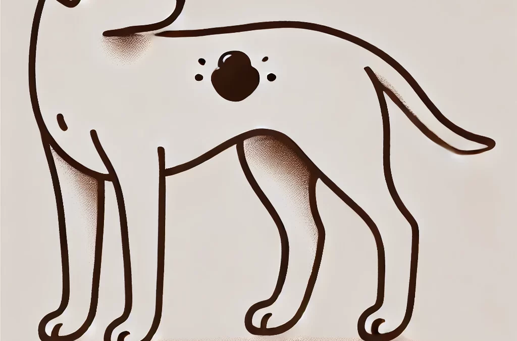
As a pet owner, discovering a lump on your dog can be alarming. The first thought that often comes to mind is, “Is it cancer?” However, not all lumps and bumps are tumors, and even if they are, not all tumors are malignant. Understanding the different types of growths, their potential causes, and the best course of action can help you make informed decisions about your dog’s health.
Common Causes of Lumps on Dogs
Lumps on dogs can arise for various reasons, ranging from benign fatty deposits to more concerning cancerous tumors. Here are some of the most common causes:
1. Lipomas (Fatty Tumors)
Lipomas are one of the most frequently found lumps on dogs, especially in older or overweight dogs. These are soft, movable, and usually harmless fatty deposits under the skin. While they typically don’t require treatment, a vet may recommend removal if they grow too large or interfere with movement.
2. Sebaceous Cysts
Sebaceous cysts occur when a hair follicle or oil gland becomes blocked, forming a lump filled with sebum (a greasy substance). These cysts can sometimes rupture and ooze a whitish or yellowish discharge. Most are benign, but they may need draining or removal if they become infected.
3. Abscesses
An abscess is a swollen, pus-filled area often caused by infections, insect bites, or wounds. These lumps can be warm, red, and painful to the touch. Abscesses may rupture on their own, but they usually require veterinary treatment, including drainage and antibiotics.
4. Warts (Papillomas)
Canine warts are caused by the papillomavirus and typically appear in younger dogs or those with weakened immune systems. These small, cauliflower-like growths usually resolve on their own but may require removal if they interfere with eating or movement.
5. Histiocytomas
Histiocytomas are benign tumors that commonly affect younger dogs. They appear as small, red, dome-shaped lumps, often on the legs, face, or ears. Many histiocytomas resolve on their own within a few months, though some may require removal if they persist.
6. Mast Cell Tumors (MCTs)
Mast cell tumors are among the most common skin cancers in dogs. They can vary in appearance—some may look like harmless lumps while others may be ulcerated or inflamed. MCTs can be aggressive, so any suspicious lump should be evaluated by a vet promptly.
7. Soft Tissue Sarcomas
These malignant tumors develop in connective tissues and can be slow-growing or aggressive. They often feel firm and may not be easily movable under the skin. Early detection and removal are crucial for a better prognosis.
How to Identify Whether a Lump is Concerning
While some lumps are harmless, others may require immediate veterinary attention. Consider the following characteristics:
- Size & Growth Rate: If a lump grows rapidly, it may indicate malignancy.
- Texture & Mobility: Soft, movable lumps are often benign, whereas firm, attached lumps can be more concerning.
- Color & Appearance: Ulcerated, inflamed, or bleeding lumps warrant immediate examination.
- Pain & Discomfort: If your dog reacts negatively to touch, it may indicate an infection or malignancy.
What to Do If You Find a Lump on Your Dog
1. Monitor the Lump
If the lump is small, soft, and not causing discomfort, you can monitor it for a few weeks. Take note of any changes in size, shape, or color.
2. Consult a Veterinarian
If the lump is growing quickly, feels firm, is painful, or has an unusual texture, schedule a vet visit. Your vet may perform a fine needle aspiration (FNA) or a biopsy to determine whether the lump is benign or malignant.
3. Consider Removal if Necessary
Benign lumps that are large, growing, or interfering with movement may need to be surgically removed. Cancerous tumors often require surgery, radiation, or chemotherapy.
4. Maintain a Healthy Lifestyle
A balanced diet, regular exercise, and routine vet check-ups can help support your dog’s immune system and overall health, reducing the risk of tumor development.
Final Thoughts
Not every lump on your dog is a cause for panic, but it’s always best to stay vigilant. Early detection and proper veterinary assessment can make all the difference in ensuring your dog’s health and well-being. If you notice any new or changing lumps, don’t hesitate to consult a veterinarian—your furry friend’s health is worth the extra attention!
Would you like additional information on any of the specific lump types or treatment options?
by TCMVET | Dec 9, 2024 | Dog Cancer & Tumors
Skin health in dogs is often a mirror of their overall well-being, yet some conditions can be puzzling for even the most attentive pet owners. One such rare condition is cornifying epitheliomas, a type of benign skin tumor that can cause concern due to its appearance and effects. Let’s delve into this unusual dermatological condition, its causes, treatments, and what makes it a unique challenge in canine healthcare.
What Are Cornifying Epitheliomas?
Cornifying epitheliomas are benign tumors that originate from sebaceous glands, specifically the epithelium (skin cells) responsible for keratin production. These tumors often present as nodular, wart-like growths on a dog’s skin. While they are not life-threatening, their potential to cause discomfort or infection means they shouldn’t be ignored.
What Causes Cornifying Epitheliomas?
The exact cause of cornifying epitheliomas isn’t fully understood, but contributing factors may include:
- Genetic Predisposition: Breeds such as Cocker Spaniels, Beagles, and Siberian Huskies are more prone to developing these growths.
- Hormonal Imbalances: Sebaceous gland activity can be influenced by hormonal changes, particularly in older dogs.
- Dietary Deficiencies: Poor nutrition can lead to imbalances in skin health, potentially exacerbating conditions like epitheliomas.
Recognizing the Symptoms
Cornifying epitheliomas typically appear as:
- Small, firm nodules with a wart-like texture
- Yellowish or waxy in color due to keratin build-up
- Localized around the head, neck, or back but can occur anywhere
- Occasionally accompanied by redness or inflammation if secondary infection occurs
While these growths are benign, rapid changes in size, color, or texture should be evaluated by a veterinarian to rule out malignancies.
Diagnosing Cornifying Epitheliomas
Diagnosis usually involves:
- Physical Examination: A veterinarian will assess the size, location, and appearance of the growths.
- Fine-Needle Aspiration (FNA): A sample of cells is extracted and analyzed to confirm the nature of the tumor.
- Biopsy: In some cases, a biopsy may be necessary to differentiate between benign epitheliomas and other skin conditions or cancers.
Treatment Options
Treatment depends on the severity and impact of the epitheliomas on your dog’s quality of life.
- Monitoring
For small, non-problematic growths, regular monitoring is often sufficient.
- Ensure the area remains clean and free from infection.
- Use soothing topical treatments if recommended by your vet.
- Surgical Removal
If the growths are causing discomfort, recurring infections, or cosmetic concerns, surgical removal is a common solution.
- Minimally invasive techniques like laser surgery can reduce recovery time.
- Topical or Systemic Therapies
- Retinoids or vitamin A supplements can regulate keratin production.
- Antibiotics may be prescribed for secondary bacterial infections.
Innovative and Natural Approaches
For owners looking to complement conventional treatments with holistic care:
- Omega-3 Fatty Acids: These can reduce inflammation and promote overall skin health.
- Herbal Remedies: Calendula and aloe vera can soothe irritated areas.
- Dietary Adjustments: A diet rich in antioxidants and high-quality proteins supports skin regeneration.
Preventive Measures
Although not all cases of cornifying epitheliomas can be prevented, these steps can help maintain optimal skin health:
- Regular Grooming: Keeps the skin clean and promotes early detection of abnormalities.
- Balanced Diet: Supports the immune system and reduces the likelihood of skin issues.
- Routine Veterinary Visits: Early intervention is key to managing any skin condition.
A Unique Challenge in Canine Dermatology
Cornifying epitheliomas highlight the importance of understanding and addressing even rare conditions in dogs. While benign, these growths can impact your pet’s comfort and appearance, making prompt and effective management essential. By staying informed and working closely with your veterinarian, you can ensure your dog remains healthy, happy, and thriving.

by TCMVET | Oct 15, 2024 | Medicines & Therapies
Lick granulomas, also known as acral lick dermatitis, are a frustrating and often chronic condition for both dogs and their owners. While they are rarely life-threatening, managing this condition can be a long-term process. Treating a lick granuloma typically requires trial and error, and several visits to the veterinarian may be necessary to find the right combination of treatments. In this article, we’ll explore effective strategies for managing lick granulomas in dogs, the importance of early intervention, and tips for preventing recurrence.
Understanding Lick Granulomas
A lick granuloma is a skin lesion caused by a dog’s compulsive licking of a particular spot, usually on the legs. Over time, this repetitive behavior leads to inflammation, infection, and thickened skin. Dogs may develop lick granulomas for various reasons, including allergies, pain, boredom, or anxiety.
Why Early Treatment Matters
Dogs with early treatment for lick granulomas tend to have a better prognosis than those with chronic or severe conditions. Left untreated, a granuloma can progress into a serious infection, potentially affecting the skin’s underlying muscles and bones. Additionally, a dog’s mouth contains harmful bacteria, making it crucial to prevent further licking to avoid worsening the condition.
Trial and Error in Treatment
Finding the right treatment plan for a lick granuloma often involves trial and error. Veterinarians may recommend a combination of treatments, including:
- Medications
Antibiotics and anti-inflammatory medications are commonly prescribed to treat infection and reduce swelling. In some cases, veterinarians may also recommend corticosteroids or antihistamines to control itching.
- Behavioral Management
Since many lick granulomas are caused by anxiety or stress, addressing the underlying behavioral issue is crucial. Calming supplements, behavioral therapy, and increased exercise or mental stimulation can help reduce a dog’s urge to lick.
- Protective Collars
One of the most effective ways to prevent further licking is by using a protective collar, such as an e-collar (Elizabethan collar). This collar should be kept on, especially when the dog is alone or at night, to ensure the granuloma is not aggravated.
- Topical Treatments
Applying topical creams or sprays that soothe the skin and deter licking may aid in healing. These treatments are often paired with antibiotics to prevent infection.
Long-Term Management of Lick Granulomas
Many dogs with lick granulomas will require long-term management rather than complete recovery. It’s important to understand that this condition can reoccur, especially if the underlying issue is not resolved. Stressful changes in your dog’s environment, such as moving homes or changes in routine, may trigger a relapse.
To manage a lick granuloma long-term, pet parents should focus on:
- Regular Vet Checkups
Regular vet visits are crucial for monitoring the condition and adjusting the treatment plan as needed.
- Environmental Enrichment
Reducing boredom and stress through environmental enrichment, such as interactive toys, regular walks, and mental challenges, can help prevent compulsive behaviors.
- Close Monitoring
Keep a close eye on any signs of recurrence. Early intervention can prevent the need for more aggressive treatments.
Lick granulomas are a challenging condition for both dogs and their owners. While they may not be life-threatening, managing the condition can require persistence and patience. Early treatment, behavioral management, and long-term care are essential to prevent further complications and help your dog live comfortably. Always consult your veterinarian for the best course of action and be prepared for ongoing care to manage this chronic condition.

by TCMVET | Oct 15, 2024 | Research and News
Acral lick granuloma, also known as acral lick dermatitis, is a common skin condition in dogs caused by excessive licking of a specific area. Typically found on the lower part of the legs, this self-induced skin lesion can lead to chronic issues if not addressed early. In this article, we’ll explore the causes, symptoms, and treatment options for acral lick granulomas, helping pet owners identify and manage this condition before it becomes a long-term problem.
What Is an Acral Lick Granuloma?
An acral lick granuloma is a skin lesion that forms due to repetitive licking of a specific area, usually the lower legs. The most commonly affected areas are the wrist (carpal joint) of the front limbs and the hock (ankle) of the back legs. Over time, the constant licking causes hair loss, redness, inflammation, and thickened skin, which can eventually lead to infection if left untreated.
Causes of Acral Lick Granulomas
Several factors can contribute to a dog’s excessive licking, leading to the formation of a granuloma. These include:
- Allergies
Allergies to food, environmental factors, or fleas can cause itchiness, prompting a dog to lick a specific area to relieve discomfort.
- Infections
Fungal, bacterial, or parasitic infections can make a dog’s skin irritated, leading to persistent licking.
- Pain
Underlying joint or bone pain, such as arthritis, may cause a dog to lick the affected area in an attempt to soothe the discomfort.
- Behavioral Issues
Stress, anxiety, or boredom can result in compulsive behaviors, including repetitive licking, which eventually leads to the formation of a granuloma.
- Neurological Issues
In some cases, nerve damage or other neurological problems can trigger excessive licking.
Symptoms of Acral Lick Granulomas
Acral lick granulomas are typically easy to spot. Some of the most common symptoms include:
- Hair loss at the licking site
- Red, inflamed skin
- Thickened or hardened skin at the affected area
- Open sores or ulcers that may become infected
- Frequent licking or chewing of the same spot
If you notice any of these signs, it’s important to consult your veterinarian for diagnosis and treatment.
Treatment Options for Acral Lick Granulomas
- Addressing Underlying Causes
The first step in treating acral lick granulomas is to identify and address the underlying cause of the licking. This may involve treating allergies, infections, or joint pain, depending on the root of the problem. Your veterinarian may prescribe antibiotics for infections, antihistamines for allergies, or pain relievers if arthritis or another pain-related issue is present.
- Behavioral Modification
If anxiety or boredom is contributing to the excessive licking, behavioral modification may be necessary. Increasing exercise, mental stimulation, and reducing stress through environmental changes can help reduce compulsive licking. Your veterinarian may also recommend anti-anxiety medications or supplements if needed.
- Topical Medications
Topical treatments, such as medicated creams or sprays, can help soothe the skin and promote healing. Some products also contain ingredients to deter the dog from licking the area further.
- Bandaging or E-Collar Use
In some cases, bandaging the affected area or using an e-collar (Elizabethan collar) can prevent further licking, giving the skin time to heal.
- Laser Therapy or Surgery
In severe or chronic cases, laser therapy or surgical removal of the granuloma may be necessary to promote healing. These treatments are typically reserved for cases that do not respond to more conservative methods.
Preventing Acral Lick Granulomas
Preventing acral lick granulomas involves addressing the potential causes of the behavior before it becomes a problem. Regular vet check-ups, maintaining a stimulating environment, and treating underlying medical issues early can reduce the likelihood of your dog developing this condition.
Acral lick granulomas are a challenging condition to treat, especially if they become chronic. Early intervention is key to preventing long-term complications, so it’s important to seek veterinary care as soon as you notice excessive licking or the development of a skin lesion. With the right combination of medical treatment, behavioral modification, and preventive care, your dog can recover and avoid further issues.

by TCMVET | Sep 23, 2024 | Dog Cancer & Tumors
Apocrine gland cysts and adenomas are common benign conditions seen in middle-aged to older dogs, particularly affecting breeds like the Great Pyrenees, Chow Chows, and Alaskan Malamutes. These cysts and adenomas develop from apocrine glands, which are associated with hair follicles and are found in the skin. This article provides an in-depth look at these conditions, including causes, symptoms, and treatment options.
What are Apocrine Gland Cysts?
Apocrine gland cysts are non-cancerous cysts that occur in the skin of dogs. They are typically seen in the middle to upper skin layers, loosely associated with hair follicles. Apocrine cysts can form on the head, neck, and other areas, often presenting as one or more fluid-filled sacs under the skin. These cysts are usually benign, meaning they do not spread or become cancerous.
There are two forms of apocrine gland cysts:
- Localized Apocrine Cysts: These develop in or near individual hair follicles and are more commonly found on the head and neck of dogs.
- Diffuse Apocrine Cysts: This form involves multiple cysts forming in the apocrine glands associated with multiple hair follicles. They can appear in areas of uninjured skin, making them harder to treat if they become widespread.
While benign, these cysts may sometimes cause discomfort, especially if they grow large or rupture.
What are Apocrine Gland Adenomas?
Apocrine gland adenomas are benign tumors that can arise in the apocrine glands. They appear as firm to soft masses and may contain fluid that ranges in color from clear to brownish. Typically, these cysts are not larger than 1.6 inches (4 centimeters) in diameter. Like apocrine cysts, adenomas are also found on the head, neck, and legs of older dogs, and, in rare cases, cats and horses.
Apocrine adenomas come in two types:
- Apocrine Adenomas: These are solid, benign growths formed by the apocrine glands.
- Apocrine Ductular Adenomas: These involve the ducts of the apocrine glands, leading to cystic formations.
Breeds at Higher Risk
Certain dog breeds are more prone to developing apocrine gland cysts and adenomas. The most commonly affected breeds include:
- Great Pyrenees
- Chow Chows
- Alaskan Malamutes
Older dogs in these breeds are more likely to develop these conditions, though the exact cause remains unknown. Genetics, environmental factors, and age may play a role in their development.
Symptoms to Look For
- Visible cysts or lumps on the head, neck, or legs
- Fluid-filled sacs under the skin
- Soft to firm masses that may vary in size
- Discoloration of the skin or cyst fluid (clear to brown)
- Possible rupture of cysts causing localized inflammation or infection
While these cysts and adenomas are generally painless, they can cause discomfort if they rupture or grow in sensitive areas.
Treatment Options
The primary treatment for apocrine gland cysts and adenomas is surgical removal. However, this can be challenging, particularly in cases where cysts are diffuse and spread across a larger area of the skin. In localized cases, the surgery is typically straightforward, and the prognosis is excellent after removal.
In situations where surgery is not an option, veterinarians may recommend monitoring the cysts for changes in size or discomfort. If cysts rupture, antibiotics may be required to prevent infection.
Apocrine gland cysts and adenomas in dogs are generally benign and treatable conditions. While they can cause cosmetic issues and occasional discomfort, early detection and treatment can help prevent complications. Owners of at-risk breeds such as Great Pyrenees, Chow Chows, and Alaskan Malamutes should monitor their dogs for the development of these cysts as they age, and consult a veterinarian for proper diagnosis and treatment.
