
by TCMVET | Jan 17, 2025 | Dog Cancer & Tumors
As a pet owner, discovering a lump on your dog can be alarming. The first thought that often comes to mind is, “Is it cancer?” However, not all lumps and bumps are tumors, and even if they are, not all tumors are malignant. Understanding the different types of growths, their potential causes, and the best course of action can help you make informed decisions about your dog’s health.
Common Causes of Lumps on Dogs
Lumps on dogs can arise for various reasons, ranging from benign fatty deposits to more concerning cancerous tumors. Here are some of the most common causes:
1. Lipomas (Fatty Tumors)
Lipomas are one of the most frequently found lumps on dogs, especially in older or overweight dogs. These are soft, movable, and usually harmless fatty deposits under the skin. While they typically don’t require treatment, a vet may recommend removal if they grow too large or interfere with movement.
2. Sebaceous Cysts
Sebaceous cysts occur when a hair follicle or oil gland becomes blocked, forming a lump filled with sebum (a greasy substance). These cysts can sometimes rupture and ooze a whitish or yellowish discharge. Most are benign, but they may need draining or removal if they become infected.
3. Abscesses
An abscess is a swollen, pus-filled area often caused by infections, insect bites, or wounds. These lumps can be warm, red, and painful to the touch. Abscesses may rupture on their own, but they usually require veterinary treatment, including drainage and antibiotics.
4. Warts (Papillomas)
Canine warts are caused by the papillomavirus and typically appear in younger dogs or those with weakened immune systems. These small, cauliflower-like growths usually resolve on their own but may require removal if they interfere with eating or movement.
5. Histiocytomas
Histiocytomas are benign tumors that commonly affect younger dogs. They appear as small, red, dome-shaped lumps, often on the legs, face, or ears. Many histiocytomas resolve on their own within a few months, though some may require removal if they persist.
6. Mast Cell Tumors (MCTs)
Mast cell tumors are among the most common skin cancers in dogs. They can vary in appearance—some may look like harmless lumps while others may be ulcerated or inflamed. MCTs can be aggressive, so any suspicious lump should be evaluated by a vet promptly.
7. Soft Tissue Sarcomas
These malignant tumors develop in connective tissues and can be slow-growing or aggressive. They often feel firm and may not be easily movable under the skin. Early detection and removal are crucial for a better prognosis.
How to Identify Whether a Lump is Concerning
While some lumps are harmless, others may require immediate veterinary attention. Consider the following characteristics:
- Size & Growth Rate: If a lump grows rapidly, it may indicate malignancy.
- Texture & Mobility: Soft, movable lumps are often benign, whereas firm, attached lumps can be more concerning.
- Color & Appearance: Ulcerated, inflamed, or bleeding lumps warrant immediate examination.
- Pain & Discomfort: If your dog reacts negatively to touch, it may indicate an infection or malignancy.
What to Do If You Find a Lump on Your Dog
1. Monitor the Lump
If the lump is small, soft, and not causing discomfort, you can monitor it for a few weeks. Take note of any changes in size, shape, or color.
2. Consult a Veterinarian
If the lump is growing quickly, feels firm, is painful, or has an unusual texture, schedule a vet visit. Your vet may perform a fine needle aspiration (FNA) or a biopsy to determine whether the lump is benign or malignant.
3. Consider Removal if Necessary
Benign lumps that are large, growing, or interfering with movement may need to be surgically removed. Cancerous tumors often require surgery, radiation, or chemotherapy.
4. Maintain a Healthy Lifestyle
A balanced diet, regular exercise, and routine vet check-ups can help support your dog’s immune system and overall health, reducing the risk of tumor development.
Final Thoughts
Not every lump on your dog is a cause for panic, but it’s always best to stay vigilant. Early detection and proper veterinary assessment can make all the difference in ensuring your dog’s health and well-being. If you notice any new or changing lumps, don’t hesitate to consult a veterinarian—your furry friend’s health is worth the extra attention!
Would you like additional information on any of the specific lump types or treatment options?

by TCMVET | Dec 14, 2024 | Dog Cancer & Tumors
When it comes to lumps on a dog’s ear, one possibility that often surprises owners is a histiocytoma. These benign growths are common in younger dogs and can appear alarming due to their rapid development and prominent location. However, they’re usually harmless and often resolve on their own. Let’s dive into what makes histiocytomas unique and how you can provide the best care for your furry friend.
What Is a Histiocytoma?
A histiocytoma is a non-cancerous tumor that arises from the Langerhans cells, a type of immune cell found in the skin. These tumors are most commonly seen in dogs under three years of age and typically occur on the head, ears, or limbs.
In the case of the ear, histiocytomas often form on the outer surface or at the edge, creating a visible lump that may concern pet parents. Despite their dramatic appearance, histiocytomas are considered one of the most benign forms of growths in dogs.
Why Do Histiocytomas Occur in Dogs?
The exact cause of histiocytomas is unknown, but they’re thought to be related to an overactive immune response in younger dogs. Breeds such as Boxers, Labradors, and Bulldogs seem to have a higher predisposition, though any breed can develop one.
How to Identify a Histiocytoma on the Ear
Histiocytomas are distinctive in their appearance and behavior, making them relatively easy to spot:
- Appearance: A raised, round, red lump, often hairless and smooth. On the ear, it can look especially pronounced due to the thin skin and lack of fur.
- Size: Typically small, around 0.5 to 2 cm in diameter.
- Texture: Firm to the touch, with a slightly ulcerated or shiny surface.
- Behavior: Unlike malignant tumors, histiocytomas don’t usually cause discomfort unless they’re scratched or infected.
Should You Be Concerned?
One of the most reassuring aspects of histiocytomas is that they’re self-limiting, meaning they often disappear without intervention within 2 to 3 months. However, veterinary consultation is crucial to confirm the diagnosis and rule out more serious conditions such as:
- Mast cell tumors
- Squamous cell carcinoma
- Ear infections or abscesses
What to Expect During Diagnosis
Veterinarians typically use the following methods to identify a histiocytoma:
- Physical Examination: A detailed inspection of the lump and overall health of the dog.
- Fine Needle Aspiration (FNA): A small sample of cells is extracted for microscopic analysis.
- Biopsy: In rare cases, a biopsy may be performed for further confirmation.
Treatment and Care Options
Most histiocytomas resolve on their own, but certain cases may require intervention:
- No Treatment Needed: If the lump isn’t bothering the dog, it can be left alone to regress naturally.
- Surgical Removal: In rare cases where the growth causes irritation or is slow to heal, a veterinarian may recommend removal.
- Topical Treatments: If the histiocytoma becomes ulcerated, antibiotic creams or sprays can prevent infection.
Home Care Tips
While waiting for the histiocytoma to resolve, there are steps you can take to ensure your dog remains comfortable:
- Prevent Scratching: Use an Elizabethan collar if your dog scratches the ear excessively, as this can lead to bleeding or infection.
- Monitor the Growth: Keep an eye on the size, color, and texture of the lump. Rapid changes may warrant a follow-up visit.
- Clean the Area: Gently clean the ear with a vet-recommended solution to reduce the risk of secondary infections.
How Is an Ear Histiocytoma Different?
Histiocytomas on the ear can be more noticeable due to the ear’s sensitive location. They may cause the ear to look asymmetrical or floppy, and dogs might shake their heads more frequently. These specific signs make monitoring and care even more important.
A Natural Approach to Supporting Your Dog’s Healing
Some pet owners explore natural therapies to complement traditional veterinary care. While these methods are not a cure, they can support overall skin health:
- Omega-3 Fatty Acids: Promote a healthy immune response and reduce inflammation.
- Calendula Cream: A soothing herbal remedy to ease irritation around the lump.
- CBD Oil: May help reduce scratching and stress associated with discomfort.
When to Seek Veterinary Advice
Even though histiocytomas are benign, consult your vet if you notice:
- The lump growing rapidly or changing in texture.
- Persistent scratching, bleeding, or discharge.
- The histiocytoma not regressing after 3 months.
Final Thoughts
A histiocytoma on your dog’s ear might seem concerning at first glance, but it’s typically a harmless condition that resolves with little to no intervention. By understanding its unique nature and providing attentive care, you can ensure your dog remains happy and healthy throughout the process.

by TCMVET | Nov 13, 2024 | Dog Cancer & Tumors
Fatty lumps, or lipomas, are one of the most common non-cancerous growths in dogs, particularly as they age. Pet owners often feel a sense of alarm when they discover these lumps, but most lipomas are benign. In this article, we’ll explore what these lumps feel like, how to distinguish them from other growths, and tips on when to consult a veterinarian.
1. What Are Fatty Lumps?
- Definition of Lipomas: Lipomas are soft, fatty deposits that form under the skin, usually in older or overweight dogs.
- Common Locations: Typically found on the torso, armpits, and limbs but can appear in various locations.
- Cause: Though exact causes aren’t fully known, they may be linked to age, breed, genetics, and weight.
2. The Feel of Fatty Lumps:
- Soft and Movable: Lipomas are generally soft and slightly squishy, feeling somewhat like a lump of dough under the skin. They’re easy to move with a gentle push, which often differentiates them from harder, less mobile growths.
- Not Painful: Unlike cysts or abscesses, lipomas are not painful unless they press on a nerve or joint.
- Consistent Texture: Lipomas usually feel the same throughout, with a homogenous, doughy texture.
3. Differences Between Lipomas and Other Types of Lumps:
- Cysts: Often firmer, cysts can be fluid-filled and sometimes have a central punctum, unlike the uniformity of a lipoma.
- Abscesses: These are usually painful and feel warm to the touch, often accompanied by redness.
- Cancerous Growths: Malignant lumps can feel harder, are often fixed in place, and can grow quickly or irregularly. Tumors might feel irregular in shape or firm.
- Fibromas: These benign tumors feel firmer than lipomas and are less mobile but are usually harmless.
4. When Should You Worry?
- Growth Rate: If a lump grows quickly, changes shape, or becomes painful, it’s best to consult a vet immediately.
- Location Concerns: Lumps near joints or in areas that affect mobility may require removal or monitoring.
- Multiple Lumps: Multiple lipomas are common in some breeds, but if new lumps appear rapidly, consider a veterinary check.
5. Diagnosing Lipomas:
- Veterinary Examination: A vet can confirm if a lump is a lipoma through palpation.
- Fine Needle Aspiration (FNA): For a more definitive diagnosis, FNA can collect cells from the lump for analysis, providing clarity on whether the lump is benign or requires further investigation.
6. Should Lipomas Be Removed?
- When Surgery is Needed: Generally, lipomas are left alone unless they cause discomfort, restrict movement, or grow excessively.
- Minimally Invasive Options: Newer methods, like liposuction or steroid injections, may help reduce lipomas in some cases.
7. Prevention and Health Tips for Reducing Lipomas:
- Maintain a Healthy Weight: Obesity is a known risk factor for lipomas.
- Regular Check-Ups: Regular vet visits allow for early detection and monitoring of any new growths.
- Balanced Diet and Exercise: A balanced diet and consistent exercise can help prevent obesity, which may reduce the likelihood of lipomas.
Conclusion: While fatty lumps in dogs can be concerning, they’re often harmless and easy to manage. By understanding the feel, causes, and symptoms of lipomas, dog owners can make informed decisions about their pet’s health. If you’re unsure about a lump on your dog, always seek professional advice for peace of mind.

by TCMVET | Sep 23, 2024 | Dog Cancer & Tumors
Metastatic tumors, or tumors that originate in one part of the body and spread to other areas, can pose a serious health risk in dogs. While it is rare for a primary tumor to metastasize to the skin, it does happen in certain cases, leading to multiple ulcerated lumps on the skin. Identifying and treating metastatic tumors is challenging, as the primary tumor is often aggressive and may spread quickly. This article explores how metastatic tumors affect dogs, their symptoms, and the importance of a whole-body treatment approach.
What Are Metastatic Tumors?
Metastatic tumors occur when cancer cells from a primary tumor elsewhere in the body spread to secondary locations, including the skin. While all malignant tumors have the potential to spread, it is uncommon for them to travel specifically to the skin. When they do, the resulting skin tumors are usually multiple and ulcerated, appearing as lumps that grow and invade deeper layers of the skin.
Why Metastatic Tumors Are Difficult to Diagnose
One of the challenges with metastatic tumors is that they may have different characteristics compared to the primary tumor. Because only a small population of cells from the primary tumor spreads to the skin, these metastatic cells may display different microscopic features, making it difficult to trace their origin. As a result, the skin tumors alone may not provide sufficient clues about the nature or location of the primary tumor.
Additionally, metastatic tumors grow quickly and often infiltrate surrounding tissue, making treatment more complicated. Early detection and identification of the primary tumor are essential for improving the chances of a positive outcome.
Symptoms of Metastatic Tumors in Dogs
- Multiple ulcerated lumps on the skin
- Lumps that grow quickly and deepen into the surrounding tissue
- Lesions that may bleed or become infected
- Signs of illness such as lethargy, loss of appetite, or weight loss, depending on the location of the primary tumor
If you notice these symptoms in your dog, it’s crucial to consult a veterinarian immediately, as metastatic tumors often spread aggressively.
Treatment for Metastatic Tumors
Because metastatic tumors involve both the skin and an internal primary tumor, treatment requires a comprehensive approach. The following are common steps in the treatment process:
- Identify the Primary Tumor: The first step is identifying the source of the metastatic tumors. This may involve advanced diagnostic tests such as imaging (X-rays, CT scans, MRIs) and biopsies.
- Treat the Primary Tumor: Once the primary tumor is identified, treatment options may include surgery, chemotherapy, or radiation therapy, depending on the tumor type and location.
- Treat the Skin Tumors: Skin tumors may require surgical removal, especially if they are causing discomfort, infection, or bleeding. However, removing the skin tumors alone is rarely sufficient if the primary tumor is not treated as well.
- Whole-Body Treatment: In many cases, systemic treatment such as chemotherapy is necessary to address both the primary tumor and any metastatic growths in the skin.
Prognosis
Unfortunately, the prognosis for dogs with metastatic tumors is often guarded to poor. Because these tumors spread rapidly and involve multiple areas of the body, achieving a complete cure can be challenging. However, early detection and aggressive treatment can improve the dog’s quality of life and, in some cases, prolong survival.
Metastatic tumors in dogs, while rare, are serious and require a whole-body approach to treatment. Recognizing the signs of these tumors, particularly ulcerated lumps on the skin, and seeking veterinary care early can make a difference in the outcome. Treatment for metastatic tumors typically involves a combination of addressing the primary tumor and managing skin growths through surgery, chemotherapy, or other therapies. Pet owners should be vigilant about monitoring their dog’s health and consulting a veterinarian if any unusual lumps or skin changes are noticed.
by TCMVET | Sep 21, 2024 | Dog Cancer & Tumors
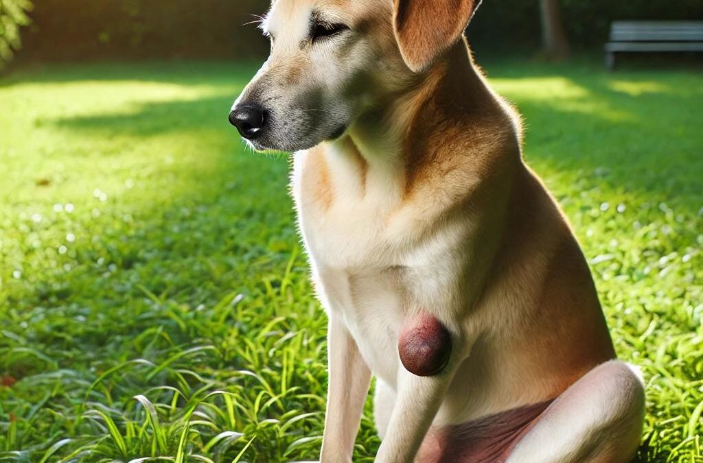
Basal cell tumors are one of the most common types of benign skin tumors found in dogs. These growths typically affect middle-aged to older dogs and can appear as firm, dome-shaped masses on the skin. While generally non-cancerous, basal cell tumors can still cause discomfort due to their size and the potential for ulceration. In this article, we will explore the symptoms, causes, and treatment options for basal cell tumors in dogs.
What Are Basal Cell Tumors?
Basal cell tumors are skin growths that originate from the basal cells found in a dog’s skin. These tumors are typically benign, meaning they are not cancerous, but they can grow large and cause discomfort. They are most commonly found on the dog’s head (especially the ears), neck, and forelimbs. Although they are benign, they may cause problems such as ulceration, inflammation, and discomfort, particularly if they grow in size.
Symptoms of Basal Cell Tumors in Dogs
Basal cell tumors generally present as firm, elevated masses on the skin. These growths may vary in size, from small bumps less than 0.4 inches (1 centimeter) to large masses over 4 inches (10 centimeters) in diameter. Some additional characteristics include:
- Solitary lumps: The tumors often appear as single masses rather than multiple growths.
- Hairless or ulcerated surface: These lumps are frequently hairless, and in some cases, they may break open and ulcerate.
- Dome-shaped growths: The masses tend to have a dome-like shape, often sticking out from the skin on stalk-like projections.
- Dark coloration: In some instances, the tumors can be dark in color.
- Secondary cysts: Cysts may form within or around the tumor.
Despite their benign nature, basal cell tumors can become problematic when they ulcerate and cause secondary inflammation. Dogs with these tumors may experience discomfort, especially if the mass becomes infected or leads to the death of surrounding skin tissue.
Causes of Basal Cell Tumors in Dogs
Several factors may contribute to the development of basal cell tumors in dogs:
- Age: These tumors are more common in middle-aged to older dogs.
- Breed predisposition: Certain breeds, including Wirehaired Pointing Griffons, Kerry Blue Terriers, and Wheaten Terriers, are more likely to develop basal cell tumors.
- Sun exposure: Prolonged exposure to UV radiation may increase the risk of skin tumors in dogs.
Treatment Options for Basal Cell Tumors in Dogs
The most effective treatment for basal cell tumors is surgical removal. Since these tumors can grow large and cause discomfort due to ulceration and inflammation, removing the tumor eliminates the source of irritation. Surgery is typically straightforward, especially when the tumor is caught early before it grows too large.
In some cases, additional treatments such as antibiotics or anti-inflammatory medications may be prescribed to manage secondary infections or inflammation caused by the tumor. Once the tumor is removed, the chances of recurrence are generally low, and the dog’s quality of life improves significantly.
Basal cell tumors in dogs, while benign, can cause significant discomfort due to their size and potential for ulceration. Regular monitoring of your dog’s skin, especially if they belong to a breed predisposed to these tumors, can help catch any growths early. Surgical removal is an effective treatment and can prevent further complications such as infections or inflammation. If you notice any unusual lumps or masses on your dog’s skin, consult a veterinarian to determine the best course of action.
