by TCMVET | Jan 20, 2025 | Dog Cancer & Tumors
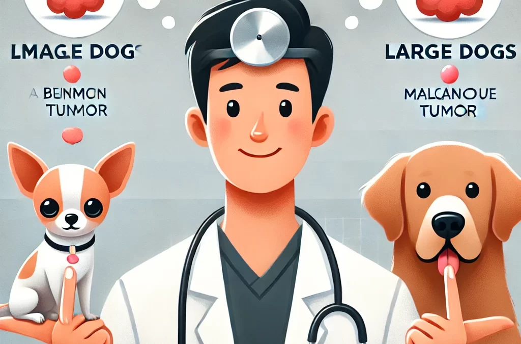
When it comes to cancer and tumor development in dogs, size matters—literally. Small and large breeds have different genetic predispositions, metabolic rates, and immune responses, all of which contribute to variations in how tumors develop, progress, and respond to treatment. If you’re a pet parent concerned about your dog’s health, understanding these differences can help you make informed decisions about prevention, early detection, and treatment.
1. The Genetic Factor: Breed-Specific Tumor Risks
Some cancers are more common in certain breeds, and a dog’s size often plays a role in this predisposition.
- Large Breeds: Dogs like Golden Retrievers, Great Danes, and Rottweilers are more likely to develop osteosarcoma (bone cancer), hemangiosarcoma (blood vessel cancer), and lymphoma. These cancers tend to be aggressive and often metastasize quickly.
- Small Breeds: Breeds like Poodles, Dachshunds, and Chihuahuas are more prone to benign tumors such as lipomas and papillomas, but they can also develop mammary tumors and bladder cancer.
The difference is not just in the type of tumors but also in how these cancers behave and respond to treatment.
2. Tumor Growth Rate and Behavior
Tumor progression varies significantly between small and large dogs due to differences in growth rates and cell metabolism.
- Faster Growth in Large Dogs: Larger breeds grow rapidly as puppies, and this rapid cell division may contribute to a higher risk of developing malignant tumors later in life. Their tumors also tend to be more aggressive.
- Slower Growth in Small Dogs: While tumors in small breeds may develop more slowly, they are still a concern. Benign tumors like lipomas are common but may interfere with mobility if they grow too large. Additionally, small dogs can still develop malignant tumors, such as mast cell tumors, which can spread if left untreated.
3. Life Expectancy and Tumor Onset
Large dogs tend to have shorter lifespans than small dogs, and this impacts tumor development timelines.
- Early-Onset Cancers in Large Dogs: Since large breeds age faster, they are more likely to develop cancer at a younger age—often between 6 to 8 years old. This means owners should begin cancer screenings and preventive care early.
- Later-Onset Tumors in Small Dogs: Small breeds may not show signs of cancer until their senior years (10+ years old), which means long-term monitoring is essential.
Understanding these timelines can help pet owners schedule vet checkups at the right time to catch potential tumors early.
4. Diagnosis and Treatment Challenges
When diagnosing and treating tumors, size plays a role in both detection and the ability to perform procedures.
- Surgical Considerations: Large dogs can better tolerate some surgeries due to their larger body mass, but removing tumors in weight-bearing bones (like with osteosarcoma) can be challenging. Small dogs, on the other hand, may struggle with anesthesia risks, especially if they are very tiny.
- Chemotherapy and Medication Differences: Dosing for chemotherapy is weight-dependent, and larger dogs often require higher drug doses, increasing treatment costs. Small dogs, despite needing lower doses, may experience stronger side effects due to their delicate systems.
5. Prevention and Early Detection Strategies
Regardless of size, early detection is crucial. Here’s what dog owners can do:
- Routine Veterinary Checkups: Regular exams help catch tumors before they grow too large.
- Physical Checks at Home: Running your hands over your dog’s body weekly can help detect unusual lumps.
- Breed-Specific Cancer Screenings: Large breeds should have early X-rays and ultrasounds, while small breeds may benefit from skin and bladder screenings.
- Diet and Lifestyle Adjustments: A balanced diet, regular exercise, and weight management can help support overall health and potentially reduce cancer risks.
Final Thoughts
While tumors affect both small and large dogs, their differences in genetics, tumor behavior, and treatment options mean pet owners need tailored care strategies. Large dogs are more prone to aggressive cancers at younger ages, while small dogs may develop slower-growing tumors later in life. By understanding these distinctions and prioritizing early detection, dog owners can improve their pets’ quality of life and potentially extend their time together.

by TCMVET | Jan 18, 2025 | Dog Cancer & Tumors
Cancer in dogs is a formidable challenge, often diagnosed too late for effective intervention. Traditional diagnostic tools such as biopsies and imaging have their limitations—they can be invasive, costly, or incapable of detecting tumors at early stages. Enter tumor biomarkers: molecular signatures found in blood, urine, or tissue that offer a game-changing approach to canine oncology. With advancements in veterinary medicine paralleling breakthroughs in human oncology, the race is on to develop reliable, non-invasive biomarkers for early detection, real-time monitoring, and personalized treatment strategies.
1. What Are Tumor Biomarkers, and Why Do They Matter?
Tumor biomarkers are measurable biological substances that indicate the presence, progression, or response to treatment of cancer. These can be:
- Proteins and Enzymes: Elevated levels of specific proteins, such as C-reactive protein (CRP) or thymidine kinase 1 (TK1), may indicate malignancies.
- Circulating Tumor DNA (ctDNA): Fragments of tumor-derived DNA found in the bloodstream offer insights into genetic mutations and tumor burden.
- Exosomes and MicroRNAs (miRNAs): Tiny extracellular vesicles and non-coding RNAs are emerging as promising tools for cancer detection and prognosis.
The ability to detect cancer before it becomes clinically evident could drastically improve treatment outcomes and quality of life for dogs.
2. The Biomarker Revolution: From Concept to Clinical Application
2.1. Early Detection: The Ultimate Game-Changer
Early-stage cancer is often asymptomatic, making routine screening a challenge. Biomarkers can fill this gap by identifying malignancies long before symptoms appear.
- Canine-Specific CRP and TK1: Elevated levels have been linked to lymphoma, hemangiosarcoma, and mast cell tumors.
- Serum microRNAs: Certain miRNA profiles correlate strongly with osteosarcoma and mammary tumors, paving the way for routine blood tests to detect high-risk cases.
2.2. Prognostic Insights: Predicting Outcomes with Precision
Not all tumors behave the same way. Biomarkers help veterinarians differentiate between aggressive cancers and slow-growing neoplasms, allowing for tailored treatment strategies.
- Ki-67 and PCNA (Proliferation Markers): High expression levels suggest rapid tumor growth and a poorer prognosis.
- LDH (Lactate Dehydrogenase): Elevated LDH levels often indicate metastasis in hemangiosarcoma, guiding treatment intensity.
2.3. Therapeutic Monitoring: Real-Time Treatment Adjustments
Biomarkers allow for non-invasive tracking of tumor response, enabling veterinarians to tweak treatments dynamically.
- Circulating Tumor DNA (ctDNA): Monitoring ctDNA levels can indicate how well a dog is responding to chemotherapy or radiation.
- Exosome Profiling: Changes in exosomal cargo composition post-treatment provide clues about residual disease and relapse risk.
3. Cutting-Edge Technologies Shaping the Future of Canine Oncology
3.1. Artificial Intelligence (AI) Meets Biomarkers
AI-powered diagnostic tools are now being trained to analyze biomarker patterns, offering near-instant, highly accurate assessments. Imagine an AI-driven blood test that predicts cancer risk before clinical signs emerge!
3.2. Liquid Biopsy: The End of Invasive Diagnostics?
Liquid biopsy, which detects ctDNA and exosomal markers, is poised to revolutionize cancer diagnostics. Unlike traditional biopsies, it offers a minimally invasive, real-time snapshot of tumor evolution.
3.3. Personalized Medicine for Dogs
As biomarker research advances, veterinarians may soon have access to biomarker-driven decision-making, selecting the best chemotherapy, immunotherapy, or targeted treatments based on a dog’s unique tumor profile.
4. Challenges and Ethical Considerations
Despite its promise, biomarker-based diagnostics face hurdles:
- Standardization Issues: Biomarker levels can vary due to breed, age, and concurrent diseases.
- Cost vs. Accessibility: Advanced biomarker tests are still expensive and not widely available.
- False Positives and Negatives: No biomarker test is 100% foolproof—further refinement is needed to ensure reliability.
5. Conclusion: The Dawn of a New Era in Canine Cancer Care
Tumor biomarkers are no longer just theoretical tools—they are rapidly becoming integral to canine cancer diagnosis, prognosis, and treatment. By embracing this molecular revolution, veterinary medicine is stepping into a future where cancer is detected earlier, treated more precisely, and monitored with unprecedented accuracy.
As technology evolves, the dream of a simple blood test that screens for multiple canine cancers could soon be a reality, giving dogs and their owners the precious gift of more time and better quality of life.

by TCMVET | Jan 18, 2025 | Dog Cancer & Tumors
The world of veterinary oncology is witnessing a dramatic shift as cutting-edge technology and visionary research converge to transform our approach to canine brain tumors. While conventional diagnostic methods and traditional treatment modalities have long guided practitioners, a new era of innovation promises to redefine clinical outcomes and enhance our understanding of these complex conditions. Below, we explore how state-of-the-art diagnostic tools, artificial intelligence, and the growing influence of stereotactic radiosurgery (SRS) are pushing the boundaries of canine neuro-oncology.
- From Symptoms to Suspicion: The Evolving Diagnostic Landscape
1.1. Recognizing the Unusual
Historically, detecting canine brain tumors hinged on recognizing subtle neurological signs—such as persistent head tilt, ataxia, and changes in behavior or appetite. While these red flags remain crucial, advanced imaging and data analytics are offering a more nuanced perspective. Practitioners are now better able to differentiate between inflammatory conditions, infections, and neoplasms thanks to high-resolution imaging and refined diagnostic algorithms.
1.2. The Rise of Advanced Imaging
• High-Field MRI: Considered the gold standard for visualizing intracranial lesions, high-field MRI provides detailed images of soft tissue and lesion boundaries. The latest MRI sequences, including functional MRI (fMRI) and diffusion tensor imaging (DTI), can delve deeper into tumor biology, revealing microstructural changes before macroscopic abnormalities appear.
• Magnetic Resonance Spectroscopy (MRS): MRS offers molecular-level insights by assessing metabolic changes within the tumor. Elevated choline and lactate peaks, for instance, can serve as early warning signs of malignancy or aggressive growth.
• AI-Assisted Image Analysis: Innovative algorithms driven by artificial intelligence are making it possible to detect and quantify tumor growth with remarkable speed and accuracy. These tools can integrate clinical data, imaging markers, and histopathological findings to predict a tumor’s likely progression or response to treatment.
1.3. Biopsy and Beyond
Though imaging technology has evolved considerably, histopathological confirmation remains a mainstay of definitive diagnosis. Stereotactic biopsy techniques minimize invasiveness, lowering complications and accelerating recovery. In the near future, liquid biopsy—analyzing circulating tumor cells or tumor DNA in the bloodstream—could further reduce the need for invasive procedures, paving the way for real-time tumor monitoring and dynamic treatment adjustments.
- The Quantum Leap: Stereotactic Radiosurgery
2.1. Breaking the Mold of Conventional Radiotherapy
For decades, external beam radiotherapy was the de facto approach to treat inoperable or surgically challenging brain tumors in dogs. While effective in certain cases, it often involved multiple sessions over several weeks. Enter Stereotactic Radiosurgery (SRS)—a precision-driven technique that delivers a concentrated radiation dose to the tumor in one or just a few sessions, minimizing damage to surrounding healthy tissue.
2.2. Hallmarks of SRS
• Pinpoint Precision: Advanced imaging and computerized treatment planning ensure the radiation beam targets only the tumor, sparing surrounding structures.
• Reduced Treatment Sessions: Many canine SRS protocols require fewer visits, thereby reducing stress for both the pet and its owner.
• Rapid Symptom Relief: High-dose radiation frequently shrinks the tumor more quickly, offering faster symptom control compared to traditional fractionated radiotherapy.
• Minimal Side Effects: The focused approach translates to fewer radiation-related complications, such as skin irritation or hair loss.
2.3. Cutting-Edge Equipment
Veterinary hospitals increasingly employ systems once reserved for human medicine, such as Gamma Knife and CyberKnife units. These devices rely on hundreds of convergent radiation beams or a robotic arm capable of delivering high-dose radiation from multiple angles—ensuring unparalleled accuracy and control.
2.4. Integrating SRS with Other Modalities
Stereotactic radiosurgery is no longer an isolated procedure. Many specialists advocate a multimodal approach, combining:
• Chemotherapy or targeted therapy to tackle microscopic disease and distant metastases.
• Immunotherapy to enhance the body’s innate ability to detect and destroy cancer cells.
• Nutritional support and rehabilitation to improve overall well-being, expedite recovery, and maintain muscle mass.
- The Road Ahead: Challenges and Opportunities
3.1. Financial and Logistical Considerations
High-end technology—such as specialized radiotherapy machines—demands significant financial outlay. Consequently, not all veterinary centers can offer SRS, limiting accessibility. However, as technology matures and more clinics adopt advanced equipment, costs may decrease.
3.2. Pushing Research Boundaries
Long-term outcome data and large-scale clinical trials remain relatively scarce in veterinary medicine. By cultivating multidisciplinary collaborations between veterinarians, oncologists, radiologists, and medical physicists, the field can gather robust evidence on the safety, efficacy, and optimal protocols for SRS in dogs.
3.3. Personalized Medicine and Beyond
Molecular profiling, genetic testing, and digital health records are merging to create a more personalized medicine environment in veterinary care. Future innovations might incorporate real-time biomarker tracking to adapt treatment strategies on the fly—potentially revolutionizing canine neuro-oncology in ways we can barely imagine today.
- Conclusion
Diagnosing and treating brain tumors in dogs has never been more sophisticated, nor more hopeful. The fusion of high-precision imaging, AI-driven analytics, and stereotactic radiosurgery is redefining what’s possible in veterinary oncology. While significant challenges remain—from ensuring widespread accessibility to gathering large-scale evidence—the collective momentum points to a brighter future for canine patients with brain tumors.
By embracing these new frontiers, we elevate not only the standard of care in canine neuro-oncology but also the broader mission of veterinary medicine itself: maximizing the health, comfort, and longevity of our beloved companions. As SRS and other groundbreaking therapies gain traction, what once seemed like a distant possibility is rapidly becoming the new norm—offering dogs with brain tumors a truly remarkable second chance at life.

by TCMVET | Dec 30, 2024 | Dog Cancer & Tumors
In recent years, the quest for holistic and integrative approaches to veterinary medicine has gained significant traction. Among these, natural therapies have emerged as promising adjuncts or alternatives in the treatment of pet tumors. As pet owners seek more holistic options to complement conventional treatments like surgery, chemotherapy, and radiation, understanding the application and efficacy of natural therapies becomes crucial. This article delves into the innovative use of natural therapies in managing pet tumors, exploring their mechanisms, benefits, and the science backing their use.
Understanding Pet Tumors
Tumors in pets, encompassing both benign and malignant forms, are increasingly common, especially in aging animals. Common types include lymphoma, mast cell tumors, osteosarcoma, and mammary tumors. Traditional treatments, while effective, often come with significant side effects and high costs. This has propelled interest in natural therapies that can potentially enhance treatment outcomes, reduce side effects, and improve the overall quality of life for pets.
Natural Therapies Explored
- Herbal MedicineHerbal remedies have been used for centuries across various cultures to treat a myriad of ailments. In the context of pet tumors, herbs like turmeric (curcumin), milk thistle (silymarin), and graviola (soursop) have shown potential anti-cancer properties. Curcumin, for instance, exhibits anti-inflammatory and antioxidant effects, potentially inhibiting tumor growth and metastasis. Milk thistle supports liver function, which is vital when pets undergo treatments like chemotherapy that can strain hepatic systems.
- Dietary InterventionsNutrition plays a pivotal role in cancer management. Diets rich in antioxidants, omega-3 fatty acids, and phytonutrients can bolster the immune system and combat oxidative stress associated with tumor progression. Incorporating foods like blueberries, kale, and fish oil into a pet’s diet may support conventional treatments and enhance overall health.
- Acupuncture and AcupressureOriginating from Traditional Chinese Medicine, acupuncture involves the insertion of fine needles into specific body points to stimulate healing. In pets with tumors, acupuncture can help manage pain, reduce inflammation, and alleviate side effects from conventional treatments. Acupressure, a needle-free alternative, offers similar benefits through the application of pressure on specific points.
- Essential Oils and AromatherapyAromatherapy employs essential oils like lavender, peppermint, and eucalyptus to promote relaxation, reduce anxiety, and improve appetite in pets undergoing cancer treatment. While not directly targeting tumors, the stress-relieving properties of aromatherapy can enhance the pet’s overall well-being and recovery process.
- Hyperthermia and CryotherapyThese therapies involve controlled application of heat or cold to tumor sites. Hyperthermia can make cancer cells more susceptible to radiation and chemotherapy, while cryotherapy can directly destroy malignant cells. When integrated with natural approaches, these therapies offer a multifaceted attack on tumor cells.
Mechanisms of Action
Natural therapies often work by targeting multiple pathways involved in tumor growth and metastasis. For example:
- Antioxidant Activity: Neutralizing free radicals to prevent DNA damage and inhibit cancer cell proliferation.
- Immune Modulation: Enhancing the pet’s immune response to recognize and destroy cancer cells more effectively.
- Anti-Inflammatory Effects: Reducing chronic inflammation that can contribute to tumor development and progression.
- Apoptosis Induction: Promoting programmed cell death in malignant cells without harming healthy tissues.
Scientific Evidence and Research
While anecdotal evidence and traditional use provide a foundation for natural therapies, scientific research is essential to validate their efficacy. Studies have shown promising results:
- Curcumin has been observed to inhibit the growth of various cancer cell lines in vitro and reduce tumor size in animal models.
- Omega-3 fatty acids have demonstrated anti-inflammatory and anti-proliferative effects in canine lymphoma.
- Acupuncture has been effective in managing chemotherapy-induced nausea and pain in pets, improving their quality of life during treatment.
However, it is crucial to approach natural therapies with scientific rigor, ensuring that treatments are evidence-based and administered safely alongside conventional therapies.
Case Studies and Success Stories
- Luna the Labrador: Diagnosed with lymphoma, Luna’s treatment included chemotherapy complemented by a turmeric-supplemented diet and acupuncture sessions. Over six months, Luna showed significant improvement in energy levels and a reduction in tumor size, highlighting the synergistic effects of combined therapies.
- Max the Maine Coon: Max was treated for a mast cell tumor with surgical removal followed by milk thistle supplements and essential oil aromatherapy. The integrated approach resulted in faster healing and minimal side effects, demonstrating the potential of natural therapies in post-surgical recovery.
Considerations and Cautions
While natural therapies offer numerous benefits, they should not replace conventional treatments but rather complement them. It is imperative to consult with a veterinarian before initiating any natural therapy to ensure compatibility with ongoing treatments and to avoid potential interactions. Dosage, quality of supplements, and individual pet health must be carefully considered to maximize benefits and minimize risks.
Conclusion
Natural therapies present a promising frontier in the management of pet tumors, offering holistic benefits that enhance conventional treatments and improve the quality of life for affected animals. As research continues to uncover the mechanisms and efficacy of these therapies, they hold the potential to revolutionize veterinary oncology. By embracing an integrative approach, pet owners and veterinarians can work together to harness the best of both natural and conventional medicine, fostering better outcomes for our beloved companions.
Future Directions
The future of natural therapies in veterinary oncology lies in comprehensive research, standardized treatment protocols, and education. Collaborative efforts between veterinarians, researchers, and holistic practitioners will be essential to establish evidence-based guidelines and optimize treatment strategies. Additionally, advancements in personalized medicine may allow for tailored natural therapy regimens that cater to the unique needs of each pet, paving the way for more effective and individualized cancer care.
Final Thoughts
The journey toward integrating natural therapies into pet tumor treatment is both exciting and challenging. By staying informed, seeking professional guidance, and prioritizing the well-being of our pets, we can navigate this evolving landscape and unlock the full potential of nature’s healing powers in veterinary medicine.

by TCMVET | Dec 26, 2024 | Dog Cancer & Tumors
A dog’s spleen is an important organ that plays a crucial role in filtering blood, storing red blood cells, and supporting the immune system. While it is not commonly discussed as much as other organs like the heart or liver, the spleen is susceptible to various health conditions, one of the most concerning being tumors. Splenic tumors in dogs can range from benign to malignant, and they can have varying implications for the animal’s health and survival.
This article aims to provide an in-depth understanding of canine splenic tumors, including the causes, symptoms, diagnosis, treatment options, and survival rates. By examining these factors in detail, we hope to provide pet owners with valuable information to make informed decisions about their dog’s health and care.
Understanding the Spleen and Its Function
Before diving into the specifics of splenic tumors, it is important to first understand the spleen’s role in a dog’s body. The spleen is located in the upper left side of the abdomen and serves several functions:
- Blood Filtration: The spleen filters and removes old or damaged red blood cells from the bloodstream.
- Storage of Blood Cells: It acts as a reservoir for red blood cells, which can be released into the bloodstream during times of increased demand, such as during physical exertion or stress.
- Immune Function: The spleen contains white blood cells that help fight infection and support the dog’s immune system.
Given these vital functions, any issue affecting the spleen can have a significant impact on a dog’s overall health.
Types of Splenic Tumors in Dogs
Splenic tumors in dogs can broadly be classified into two categories: benign and malignant. Understanding the distinction between these two types is crucial in determining the prognosis and treatment approach.
- Benign Tumors
- Hemangiomas: These are tumors made up of blood vessels and are considered benign. Hemangiomas are often small and may not cause noticeable symptoms unless they grow large enough to interfere with the spleen’s function.
- Benign Nodular Hyperplasia: This is a non-cancerous growth of spleen tissue, typically seen in older dogs. It is often asymptomatic and may not require treatment unless it becomes problematic.
- Malignant Tumors
- Hemangiosarcoma: This is the most common malignant splenic tumor in dogs and is known for its aggressive nature. Hemangiosarcoma arises from blood vessel cells and can spread rapidly to other organs such as the liver, lungs, and heart.
- Lymphoma: This type of cancer originates in the lymphatic system and can affect the spleen, leading to a condition called splenic lymphoma. Lymphoma is typically associated with systemic disease and can affect other parts of the body as well.
- Leukemia: Although rare, leukemia can involve the spleen, and it results in an abnormal increase of white blood cells that may lead to splenomegaly (enlarged spleen) and other symptoms.
The prognosis for dogs diagnosed with malignant splenic tumors, particularly hemangiosarcoma, tends to be poor due to the rapid progression of the disease.
Symptoms of Splenic Tumors in Dogs
The symptoms of splenic tumors can be vague and may mimic those of other abdominal conditions. Some dogs with splenic tumors may not show any obvious signs of illness, while others may experience severe symptoms as the tumor grows or ruptures.
Common symptoms include:
- Abdominal Distension or Swelling: This can occur if the tumor grows large enough to cause the spleen to become enlarged.
- Lethargy or Weakness: Dogs with splenic tumors often become tired easily and may exhibit a lack of energy or interest in their usual activities.
- Pale Gums: If the tumor causes internal bleeding, a dog may develop pale gums, which is a sign of anemia.
- Collapse or Sudden Death: This can occur if the tumor ruptures, leading to internal bleeding. Hemangiosarcoma, in particular, is known for causing sudden internal bleeding that can be fatal if not treated immediately.
- Loss of Appetite and Weight Loss: Dogs with splenic tumors may refuse food and experience unexplained weight loss.
- Vomiting or Diarrhea: Digestive disturbances are also common in dogs with abdominal tumors.
If a dog exhibits any of these symptoms, it is important to seek veterinary care as soon as possible to rule out or confirm the presence of a splenic tumor.
Diagnosing Splenic Tumors in Dogs
The diagnosis of a splenic tumor usually involves a combination of physical examination, imaging tests, and sometimes biopsies. The steps typically include:
- Physical Examination: The veterinarian will palpate the dog’s abdomen to check for signs of an enlarged spleen or abdominal tenderness.
- Blood Tests: Blood work may show signs of anemia or other abnormalities, such as a low platelet count, which may suggest internal bleeding.
- Imaging Tests: X-rays, ultrasound, or CT scans are used to visualize the spleen and other abdominal organs. An ultrasound, in particular, is useful for identifying the size, shape, and location of the tumor.
- Fine Needle Aspiration (FNA): A fine needle aspiration involves extracting a small sample of tissue from the spleen to evaluate for cancerous cells. This is often done if the tumor is visible on an ultrasound.
- Surgical Exploration: In some cases, surgery may be performed to remove the spleen or part of it for biopsy, especially if other diagnostic tests are inconclusive.
The most common malignant splenic tumor in dogs, hemangiosarcoma, is often diagnosed after it has already caused significant damage, such as internal bleeding or metastasis to other organs.
Treatment Options for Canine Splenic Tumors
The treatment for splenic tumors in dogs largely depends on the type of tumor, its stage, and whether it has metastasized. The main treatment options include surgery, chemotherapy, and supportive care.
- Surgical Removal (Splenectomy): If the tumor is localized to the spleen and has not spread to other organs, surgery to remove the spleen (splenectomy) is typically the first line of treatment. Splenectomy is generally well-tolerated by dogs, and they can live without a spleen, as other organs (such as the liver) take over its functions.
- Prognosis After Surgery: For benign tumors or if the malignancy is detected early, the prognosis after surgery can be good. However, for tumors like hemangiosarcoma, the prognosis remains poor, even after the spleen is removed.
- Chemotherapy: If a malignant tumor like hemangiosarcoma has spread to other organs or is considered high-risk, chemotherapy may be recommended after surgery to help manage the disease. While chemotherapy can extend survival time, it is often not curative.
- Chemotherapy Protocols: Common chemotherapy drugs used for hemangiosarcoma include doxorubicin, cyclophosphamide, and vincristine. Treatment plans may vary depending on the individual dog’s condition.
- Supportive Care: Dogs that are diagnosed with splenic tumors may require supportive care to manage symptoms and improve quality of life. This may include:
- Blood transfusions for anemia or blood loss
- Pain management, especially in cases of internal bleeding or discomfort
- Nutritional support if the dog is not eating properly
In cases where surgery or chemotherapy is not an option, palliative care may help improve the dog’s comfort.
Survival Rates and Prognosis
The prognosis for dogs with splenic tumors varies significantly depending on several factors, including the type of tumor, its stage, and the success of treatment.
- Benign Tumors: For dogs with benign splenic tumors, such as hemangiomas or benign nodular hyperplasia, the prognosis is generally good, especially if the tumor is detected early and surgically removed. These dogs can live normal, healthy lives post-surgery.
- Malignant Tumors:
- Hemangiosarcoma: This aggressive cancer is notorious for its poor prognosis. The survival rate for dogs with hemangiosarcoma, even with surgery to remove the spleen, is typically only 3 to 6 months without chemotherapy. With chemotherapy, survival can be extended to 6 to 12 months, but the cancer often recurs.
- Lymphoma and Leukemia: The prognosis for splenic lymphoma and leukemia depends on the type and stage of the disease, as well as the dog’s response to treatment. Chemotherapy can help control the disease, but these conditions can also be fatal if not managed effectively.
Conclusion
Splenic tumors in dogs present a significant health challenge, particularly when they are malignant. The prognosis varies widely depending on the type of tumor and the stage at diagnosis. While benign tumors have a relatively favorable outcome with surgical removal, malignant tumors such as hemangiosarcoma often have a poor prognosis, with treatment focused on extending life and managing symptoms.
For pet owners, early detection is key. If you notice any signs of illness or unusual behavior in your dog, it is important to consult a veterinarian promptly. With proper diagnosis and treatment, many dogs with splenic tumors can lead fulfilling lives, and in some cases, their chances of survival can be significantly improved.
Understanding the symptoms, treatment options, and survival rates of splenic tumors is essential for responsible pet ownership and ensuring the best possible care for your dog.
